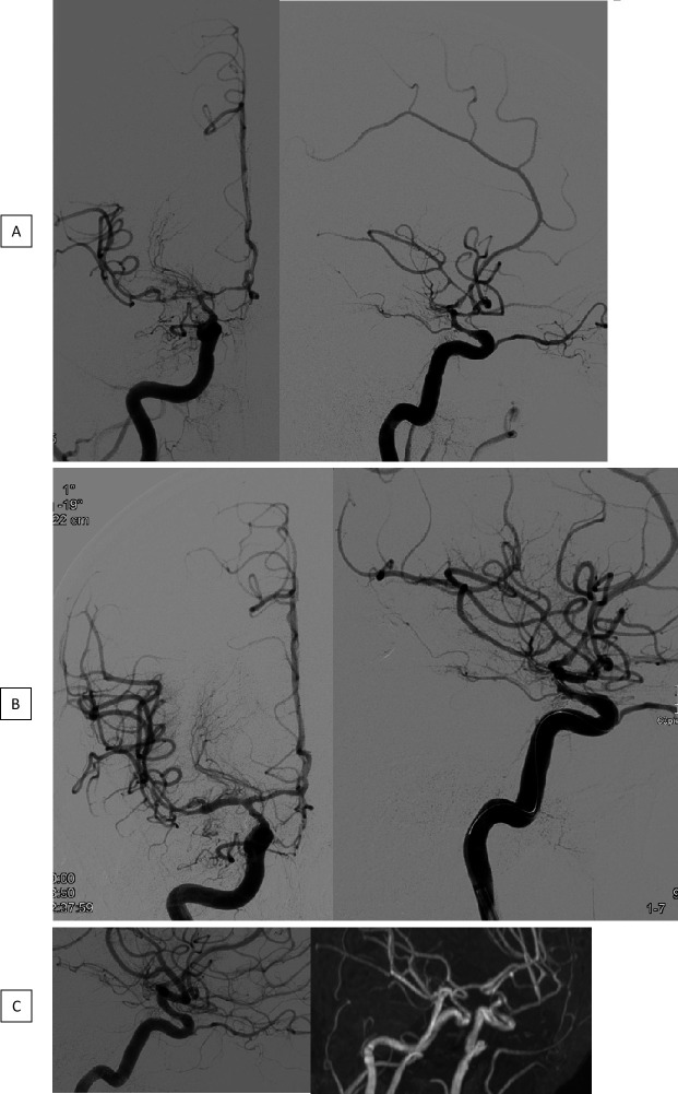
Figure 2.
Angiogram before (A) and after (B) TBA with a CB in connection with severe CVS in the distal ICA as well as within the proximal segments of the MCA and ACA. Mild arterial narrowing in the catheter angiography and TOF MRA, 6 months later (C). ACA, anterior cerebral artery; CB, compliant balloons; CVS, cerebral vasospasm; ICA, internal carotid artery; MCA, middle cerebral artery; TBA, transluminal balloon angioplasty; TOF MRA, time-of-flight magnetic resonance angiography.

