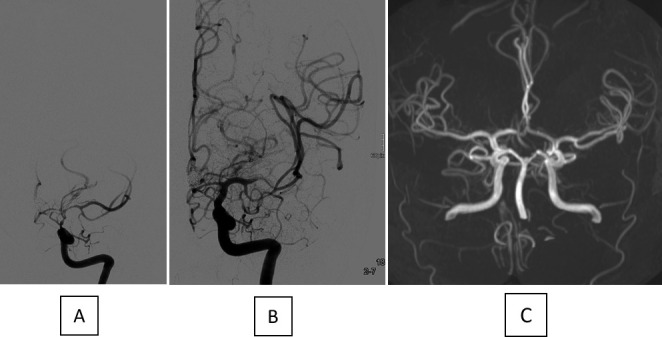
Figure 3.

Angiogram before (A) and after (B) TBA with a CB in connection with severe CVS in the distal ICA as well as the proximal segments of the MCA and ACA. No arterial narrowing is visible in the TOF MRA 11 months later (C). ACA, anterior cerebral artery; CB, compliant balloon; CVS, cerebral vasospasm; ICA, internal carotid artery; MCA, middle cerebral artery; TBA; transluminal balloon angioplasty; TOF MRA, time-of-flight magnetic resonance angiography.
