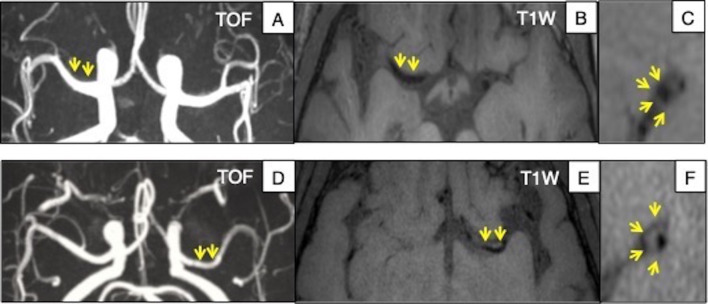
Figure 1.
Example of intracranial MRI in USA and China. Upper row: Caucasian subject, (A) TOF MIP image shows no significant stenosis in the MCA. (B) and (C) Axial and sagittal views of 3D-T1 VISTA image shows no wall thickness. Bottom row: Chinese subject, (D) TOF MIP image shows no significant stenosis in the MCA. (E) and (F) Axial and sagittal views of 3D-T1 SPACE image reveals extraluminal hyperintensity of MCA segment, indicating plaque without significant stenosis. MCA, middle cerebral artery; MIP, maximum intensity projection.

