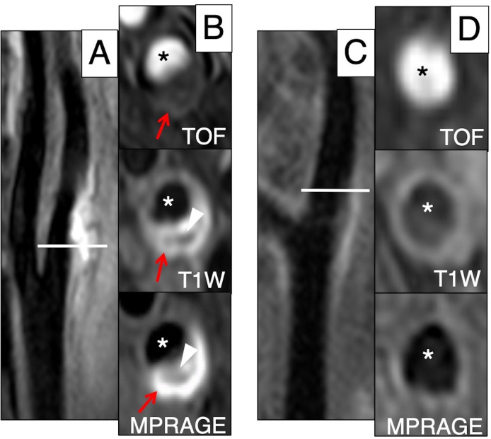
Figure 2.
Example of extracranial carotid MRI in USA and China. Left: Caucasian subject, (A) Longitudinal view of extracranial carotid artery. The horizontal line indicates the level of the transverse carotid plaque images (shown in B). (B) The high-intensity area on TOF, T1W and MPRAGE indicates regions of IPH (red arrow). The low-intensity area on T1W and MPRAGE indicates a LRNC (arrowhead). Right: Chinese subject, (C) longitudinal view of extracranial carotid artery. The horizontal line indicates the level of the transverse carotid plaque images (shown in D). (D) TOF shows no significant stenosis and T1W and MPRAGE show no plaque. Asterisk represents lumen. IPH, intraplaque haemorrhage; LRNC, lipid-rich necrotic core.

