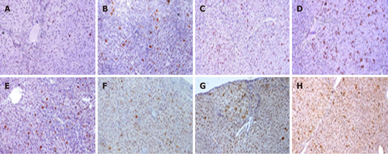
Figure 4.
Images of rats' liver sections stained immunohistochemically with proliferating cell nuclear antigen. Positive immune-reactive nucleus (brown dots) scatter in-between negatively stained liver tissue of rats who received diethylnitrosamine (DEN) and different doses of 2-acetylaminofluorene (2-AAF). A-D: Rats sacrificed at week 10; E-H: Rats sacrificed at week 16 (A and E: DEN group; B and F: DEN + 100 mg 2AAF group; C and G: DEN + 200 mg 2AAF group; D and H: DEN + 300 mg 2AAF; magnification × 100).

