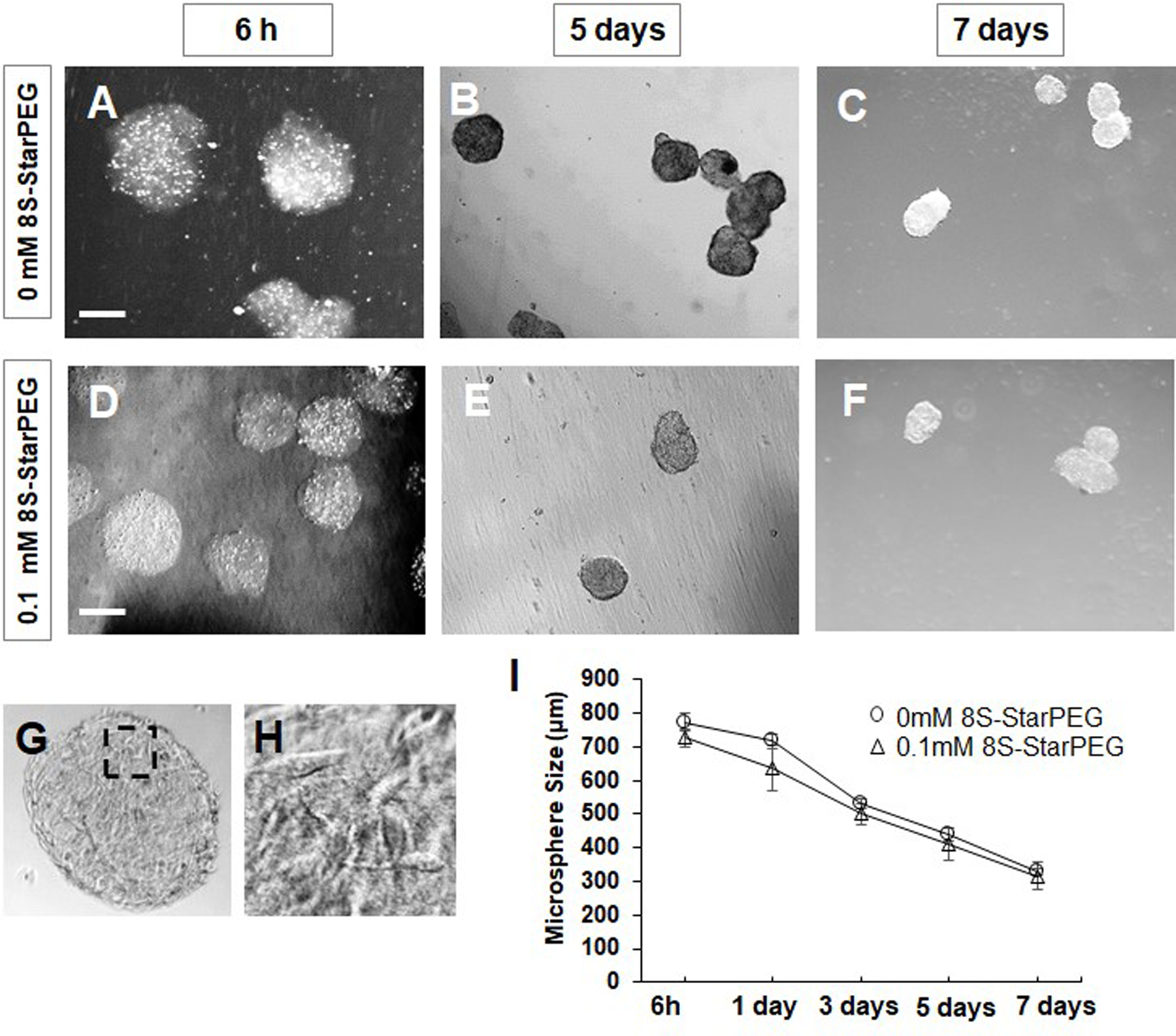
Figure 1.

Fabrication of microtumors of U87 glioma cells. (A–C) Non-crosslinked microtumors and (D–F) Microtumors crosslinked with 8S-StarPEG and cultured for 7 days in cell culture medium. Microtumor size decreased gradually. Scale bar: 400 µm. (G) Dense tumor cells seen in microtumors after 7 days. (H) Magnified image of inset indicated in (G). (I) Quantification of microtumor size showing decrease when cultured in cell culture medium.
