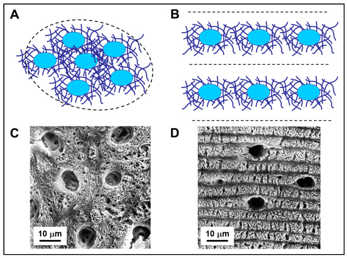
Figure 4.
(A,B): Schematic drawing showing arrangement of osteocytes and the surrounding collagen fibers in not-lamellar-woven-bone (A) and in lamellar-woven-bone (B). In both types of bone tissue, osteocyte lacunae (blue ovals) are surrounded by perilacunar matrices of loosely arranged collagen fibers. In lamellar-woven-bone, the loose lamellae result as a consequence of alignment and fusion of the perilacunar loose matrices of the osteocytes arranged in planes; the dense bundles of collagen fibers do not contain osteocytes. (C,D): SEM micrographs of not-lamellar-woven-bone (C) and lamellar-woven-bone (D); the osteocyte lacunae are larger, more numerous, and irregularly distributed in not-lamellar-woven-bone with respect to lamellar-woven-bone, where they are only located inside loose lamellae. Note that dense lamellae, alternating with the loose ones, are thinner and do not contain osteocyte lacunae.

