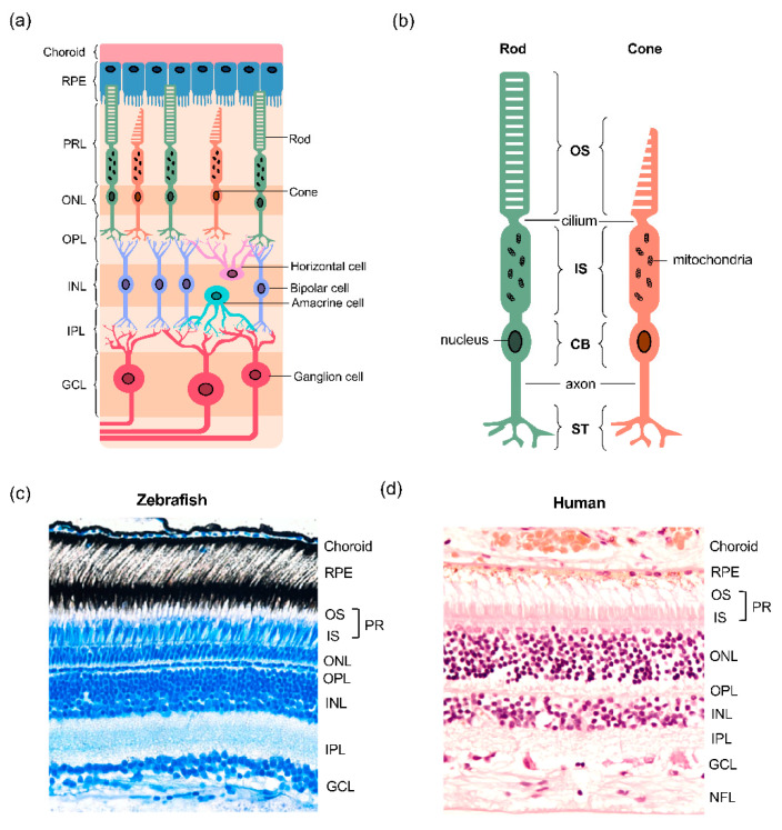
Figure 1.
Structural features of the vertebrate retina. (a) Overview of the multicellular layered retina. The retinal pigment epithelium (RPE) sits between the photoreceptor layer (PRL) in the outer retina and the choroidal vasculature. The cell bodies of rod and cone photoreceptors are located in the outer nuclear layer (ONL) and form synapses with horizontal and bipolar cells in the outer plexiform layer (OPL). The inner nuclear layer (INL) contains the cell bodies of horizontal, bipolar and amacrine cells, the latter two forming synapses with retinal ganglion cells (RGC) in the inner plexiform layer (IPL); (b) structure of the rod and cone photoreceptors. Both photoreceptor types have a light-sensitive outer segment (OS) connected to the inner segment (IS) by a single connecting cilium. The IS is abundant in megamitochondria, an adaptation that maximises energy production. The cell body contains the nucleus and protein synthesis machinery, and the axon separates the cell body (CB) from the synaptic terminals (ST), whilst transmitting the action potential that triggers glutamate neurotransmission release from ST; (c,d) a comparison of the structure of the human and zebrafish retinae.

