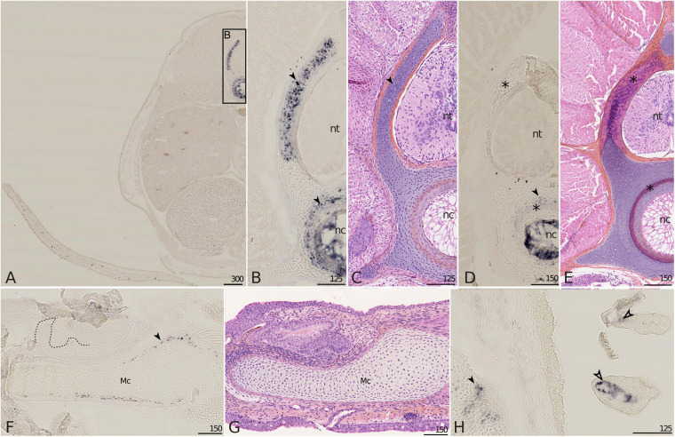
FIGURE 5.
Mgp1 gene expression on sections of late developing embryos of the small-spotted catshark Scyliorhinus canicula. (A–C) A total of 6 cm long embryos showing Mgp1 in situ hybridization, general (A) and closer (B) view on transverse sections at the level of the pectoral fin and Hematoxylin-Eosin-Saffron (HES) staining of a comparable zone to B (C). (D,E,H) Transverse sections of 7.7 cm long embryos displaying Mgp1 in situ hybridization (D,H) or HES staining (E). (F) Mgp1 in situ hybridization on a parasagittal section of the Meckel’s cartilage of a hatchling embryo with developing teeth [dotted line separates the epithelial (e) and mesenchymal (m) compartments of teeth]. (G) HES staining on a comparable zone to (F). (H) Branchial basket with gills. Mgp1 expression is detected in neural arch and vertebral body chondrocytes (filled arrowheads in B,D) before but not after mineralization (located with asterisks in D,E); in chondrocytes in the periphery of the Meckel’s cartilage before mineralization (filled arrowhead in F) and of other skeletal elements (filled arrowhead in H); in the connective tissue cells that surround vasculature in gills (open arrowhead in H). Mc, Meckel’s cartilage; nc, notochord; nt, neural tube. Scales are in μm.

