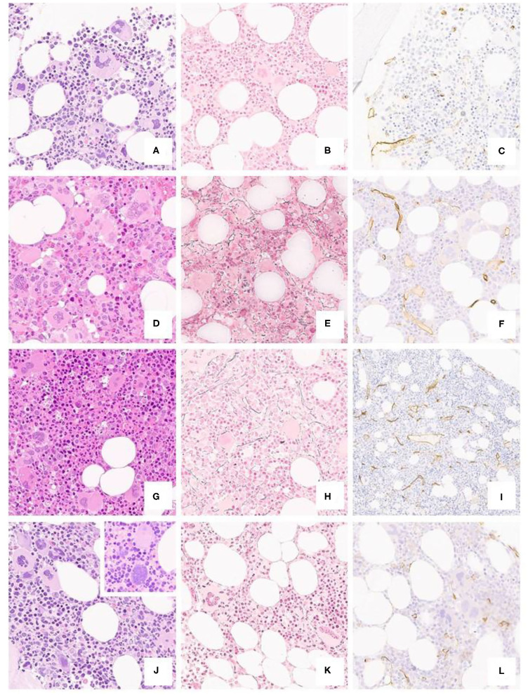
Figure 2.
Typical ET case from a 40-year-old patient (a H/E, 20x), featuring a polymorphic spectrum of megakaryocytes, but with giant, hyperlobated forms and scant naked nuclei, MF-0 fibrosis (b Gomori, 20x) and unremarkable CD34+ blasts and vessels (c CD34, 20x). BM biopsy from a 35-year-old subject (d H/E, 20x) featuring mild hypercellularity, increased, left-shifted erythropoiesis with proerythroblasts and absolute predominance of giant, hyperlobated megakaryocytes; there is only a minor increase in reticulin fibers (e Gomori, 20x) and vessel density (f CD34, 20x). A further case from a 31-year-old patient shows mild panmyelosis (g H/E, 20x) with polymorphic megakaryocyte clusters, but featuring ET-like giant forms, along with dim reticulin fibers increase (h Gomori, 20x) and mild micro-vessel proliferation (i CD34, 20x). The last panel from a 52-year-old individual depicts a normocellular bone marrow (j H/E, 20x) with clustering of predominantly hyperlobulated megakaryocytes, with scant bulbous forms (inset) or with maturation defects, with unremarkable fibrosis and CD34+ positive cells (k Gomori, 20x; l CD34, 20x).

