
Figure 6.
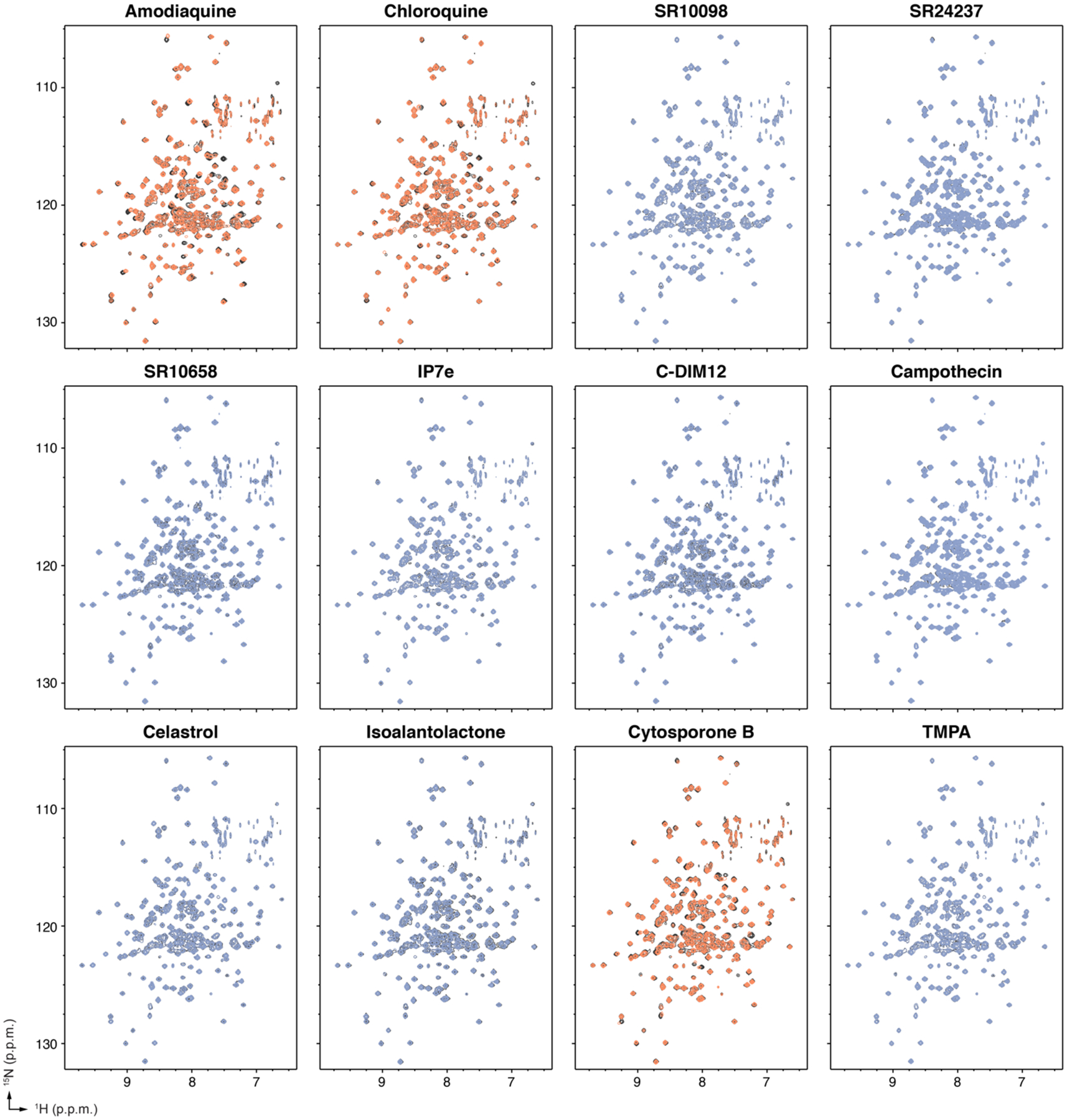
Protein NMR spectroscopy ligand footprinting data. 2D [1H,15N]-TROSY-HSQC data (700 MHz, 298K) of 15N-labeled Nurr1 LBD in the presence of vehicle control (black spectra) or 2X ligand reveals ligands that do not bind to the Nurr1 LBD (blue spectra) and ligands that bind to the Nurr1 LBD (orange spectra).