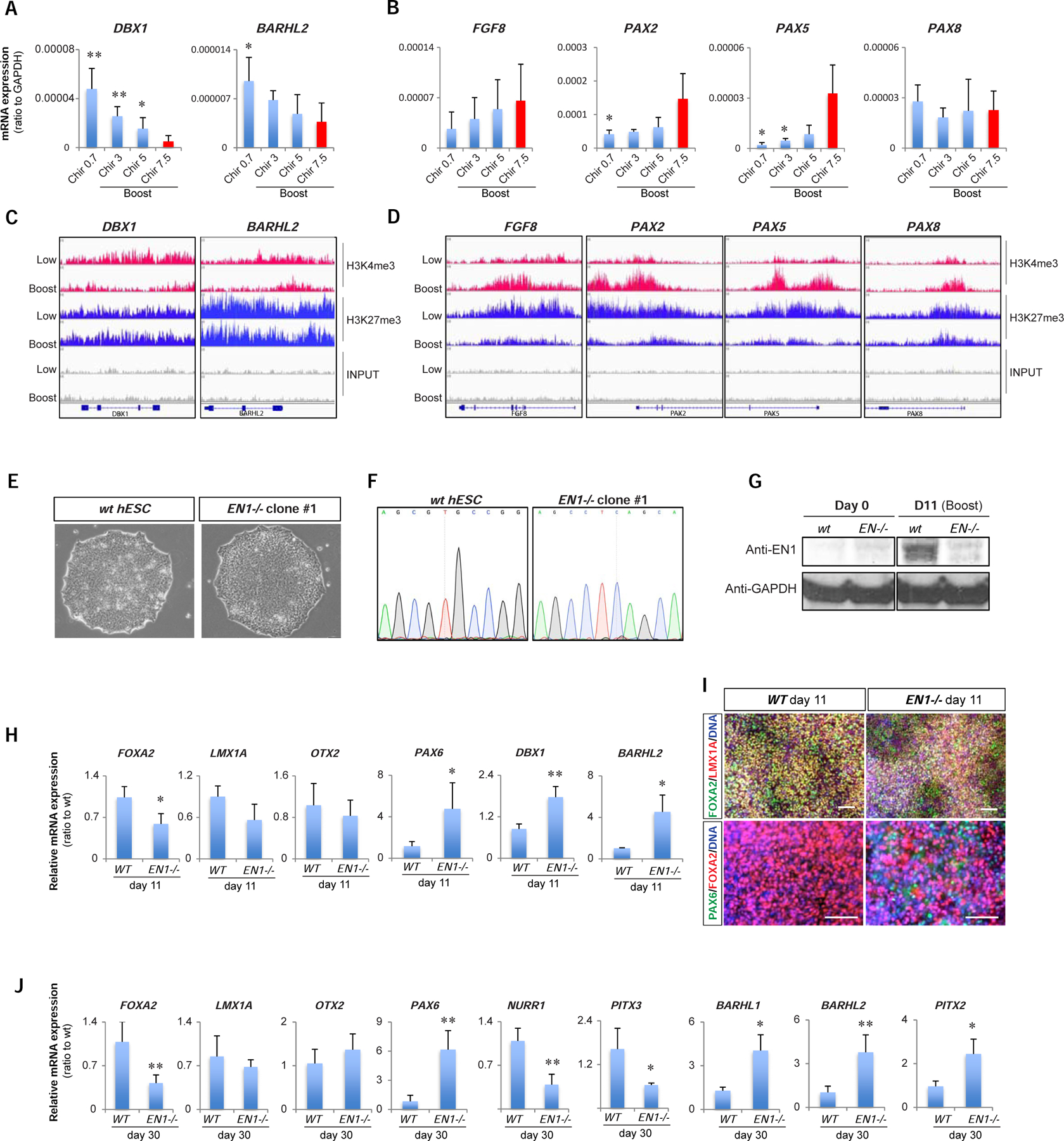
Figure 2. Suppression of subthalamic fate in Boost CHIR is dependent on EN1.

(A, B) qRT-PCR of subthalamic nucleus markers (DBX1 and BARHL2; A) and FGF8 related genes (FGF8, PAX2, PAX5, and PAX8; B) among different CHIR-boost treated cells at day 11. (C-D) IGV view of ChIP-sequencing data for H3K4me3 and H3K27me3 between Low- and Boost-CHIR treated mDA differentiated cells at day 11 at the loci of subthalamic nucleus markers (DBX1 and BARHL2; C) and FGF8 related genes (FGF8, PAX2, PAX5, and PAX8; D). (E) hPSC morphology of wild-type (WT) and EN1 knockout (EN1−/−) clones. (F) Sanger sequencing chromatograms comparing WT and EN1−/− hPSC clones. (G) Western-blotting of EN1 and GAPDH between day 0 and day 11 mDA differentiated cells from WT and EN1−/− hPSCs. (H, I) qRT-PCR analysis (H) and immunofluorescent staining (I) of day 11 mDA differentiated cells from WT and EN1−/− hPSCs. Scale bars = 100 µm. (J) qRT-PCR analysis of day 30 of mDA neuron differentiation from WT and EN1−/− hPSCs.
