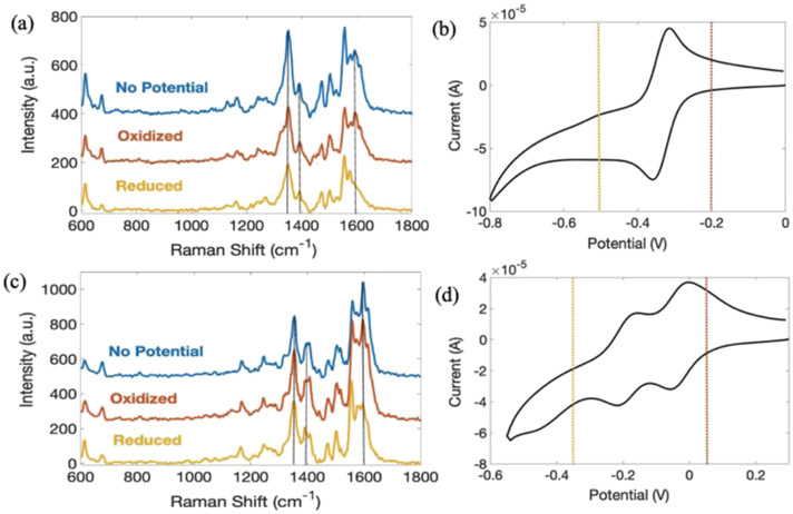
Figure 2.
SERS spectra of PYO at pH 7 (a) and pH 2.4 (c) as a function of applied potential and corresponding CV of PYO at pH 7 (b) and pH 2.4 (d). All spectra were obtained at an integration time of 0.5 s with 10 accumulations, and potentials were applied for 20 s. At pH 7, oxidizing potential, red dashed line in (b), Eox = −0.2 V, and reducing potential, yellow dashed line in (b), Ered = −0.5 V, both vs Ag/AgCl, were applied. At pH 2.4, oxidizing potential, red dashed line in (d), Eox = 0.05 V and reducing potential, yellow dashed line in (d), Ered = −0.35 V, both vs Ag/AgCl, were applied.

