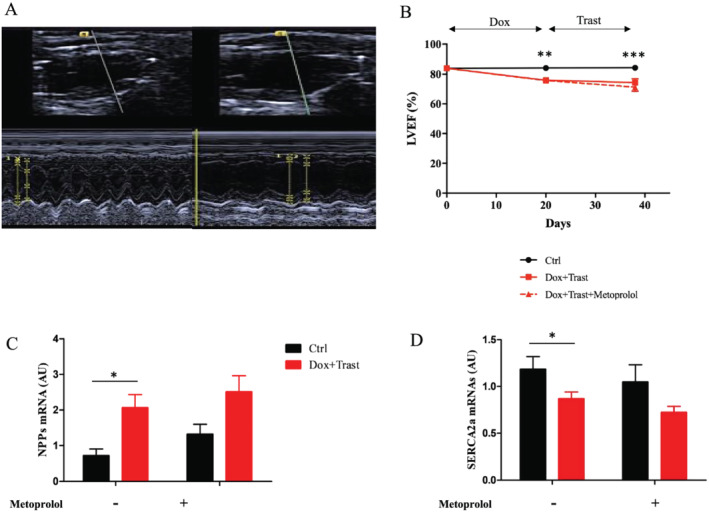
Figure 2.

Effects of metoprolol on chemotherapy‐induced cardiac dysfunction. (A) Typical echo image of LV dysfunction without any LV dilation at Day 38 after chemotherapies were initiated. (B) The measure of LVEF with echocardiography from baseline to Day 38 indicates a rapid decline in LVEF, which is not prevented by metoprolol treatment (Ctrl with and without Meto n = 15, Dox + Trast n = 11, Dox + Trast + Meto n = 8, **P < 0.01, ***P < 0.001 by two‐way using ANOVA test followed by Bonferroni post hoc test). (C–D) Quantification of natriuretic peptides (NPPs) (C) and SERCA2a (D) mRNAs in ventricular tissues. Note the significant increase of NPPs after chemotherapy. The decrease in SERCA2a mRNA level is a sign of heart failure. Values are means ± SEM, *P < 0.05, **P < 0.01 by two‐way using ANOVA test followed by Bonferroni post hoc test. (Ctrl n = 8; Ctrl + Meto n = 5, Dox + Trast n = 10, Dox + Trast + Meto n = 7).
