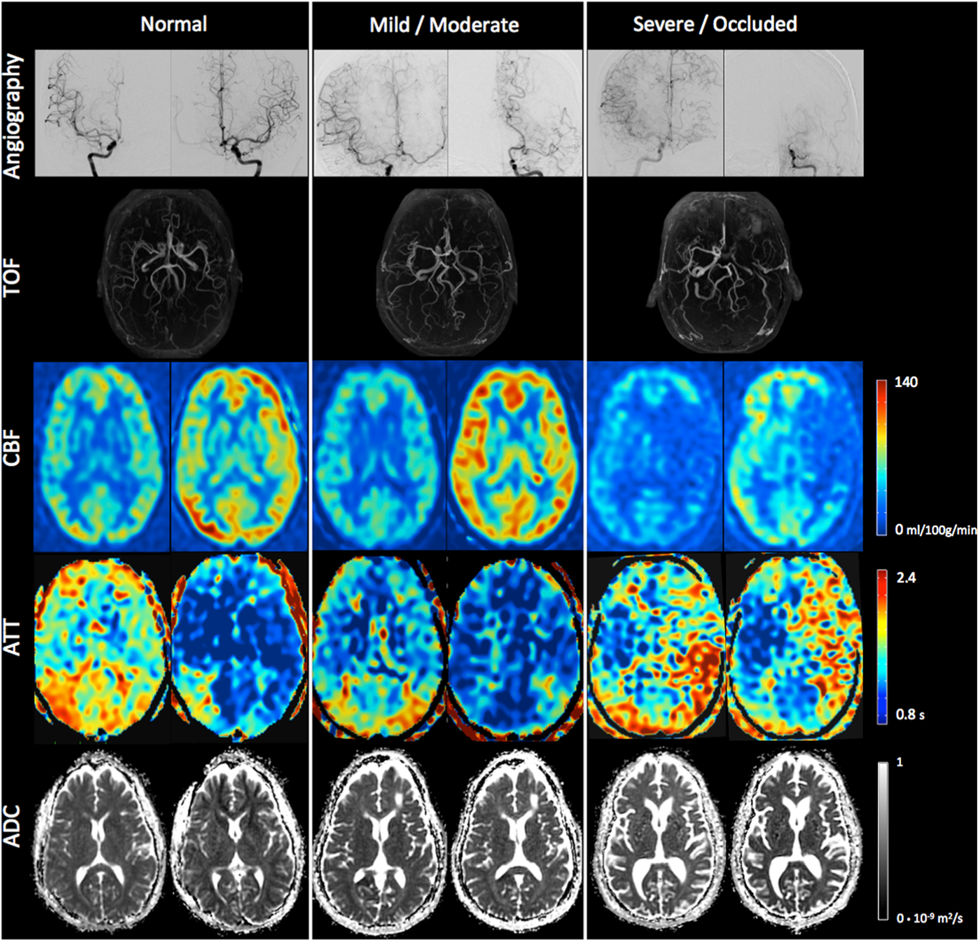
Fig. 2.

(Top) Catheter angiography (right and left internal carotid injection) and TOF. (Bottom) CBF, ATT, and ADC at baseline (left) and after acetazolamide challenge (right) in patients with normal, mild/moderate, and occluded left MCA, respectively. All of the other territories were without arterial stenosis in those three patients. A significant CBF increase is visible in all territories after acetazolamide, except in the severely stenosed/occluded left MCA. A decrease in ATT is visible in all territories after acetazolamide. Interestingly, slightly prolonged ATT is visible in the posterior part of the right MCA in the patient with normal vessels on the MR angiogram, suggesting the ASL ATT measurement is very sensitive, and can depict even mild delays
