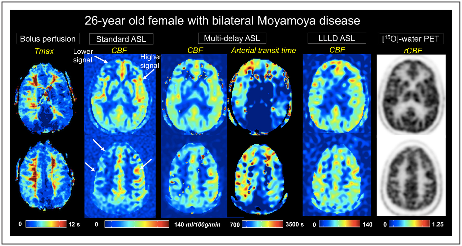
Figure 1.

A 26-year-old female patient with bilateral Moyamoya disease. Time-to-maximum (Tmax) images from dynamic susceptibility contrast MRI are shown on the leftmost panel. Cerebral blood flow (CBF) maps (mL/100 g per minute) from each of the 3 arterial spin labeling (ASL) acquisitions are depicted at 2 slice locations, as well as arterial transit time from multidelay ASL. Arrows indicate areas of lower and higher signal on standard single-delay ASL relative to the [15O]-water positron emission tomography (PET) reference (far right). Improved cortical CBF measurement compared with PET is seen on multidelay and long-label long-delay (LLLD) ASL scans. rCBF indicates relative CBF.
