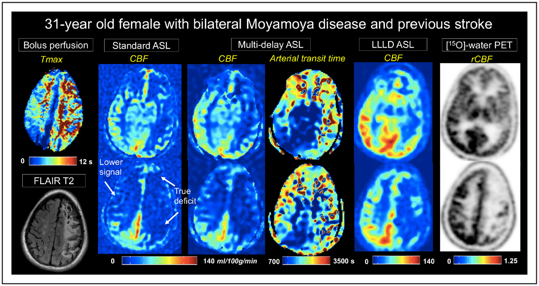
Figure 2.

A 31-year-old women with severe, bilateral Moyamoya disease and a previous stroke on left hemisphere. Time-to-maximum (Tmax) images from dynamic susceptibility contrast are shown on the leftmost panel. Cerebral blood flow (CBF) maps (mL/100 g per minute) from each of the 3 arterial spin labeling (ASL) acquisitions are depicted at 2 slice locations, as well as arterial transit time from multidelay ASL. Signal loss on standard single-delay ASL relative to positron emission tomography (PET), because of long arterial transit times, are evident in the right hemisphere. This signal loss is partially recovered in multidelay ASL, and more fully recovered with long-label long-delay (LLLD) ASL. LLLD ASL allows distinction between nonperfused tissue from the stroke and perfused tissue with extremely long Tmax. rCBF indicates relative CBF.
