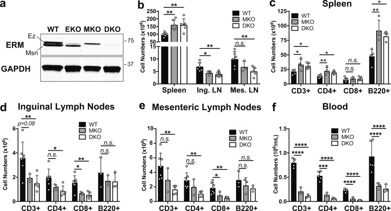
Figure 1.
ERM-deficient lymphocytes assume an altered tissue distribution with severe lymphopenia. (a) Lysates from CD4+ T cell blasts were immunoblotted with a pan-ERM antibody and anti-GAPDH. (b) Total cell counts from spleens (after RBC lysis), inguinal lymph nodes (Ing. LN), and mesenteric lymph nodes (Mes. LN) from 8–16-wk-old mice. (c–f) Cell suspensions and peripheral blood mononuclear cells were analyzed by flow cytometry. (b–f) Data represent means ± SD. Each dot represents an individual mouse; n = 3–7 mice per group. Statistical significance was determined using a standard one-way ANOVA. Data distribution was assumed to be normal, but this was not formally tested. Ez, ezrin; Msn, moesin. n.s., P > 0.05; *, P < 0.05; **, P < 0.01; ***, P < 0.001; ****, P < 0.0001.

