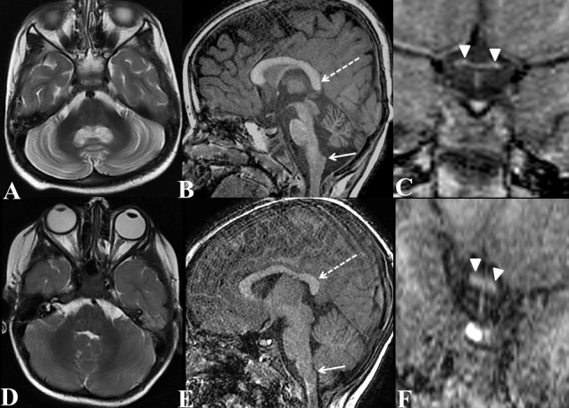
Figure 1.

Axial T2W (A) of brain, sagittal T1W of brain (B) and coronal T1W image of optic chiasma (C) of our patient with homozygous p.Gly586Arg variant in PLA2G6; compared with axial T2W (D) of brain, sagittal T1W of brain (E) and coronal T1W image of optic chiasma (D) of a normal 3-year-old boy (age and gender matched normal subject). Axial T2W image (A) shows diffuse symmetrical atrophy with gliosis (hyperintensities) of bilateral cerebellar hemispheres (compare with normal cerebellum of normal child of same age in D). In addition, sagittal T1W image of brain (B) shows claval (nucleus gracilis) hypertrophy (arrow) with drooping of splenium of corpus callosum (broken arrow), (compare with normal claval and splenium of corpus callosum of normal child of same age in E). Coronal T1W image of optic chiasma (C) shows diffuse optic chiasma atrophy (compare with normal optic chiasma of normal child of same age in F).
