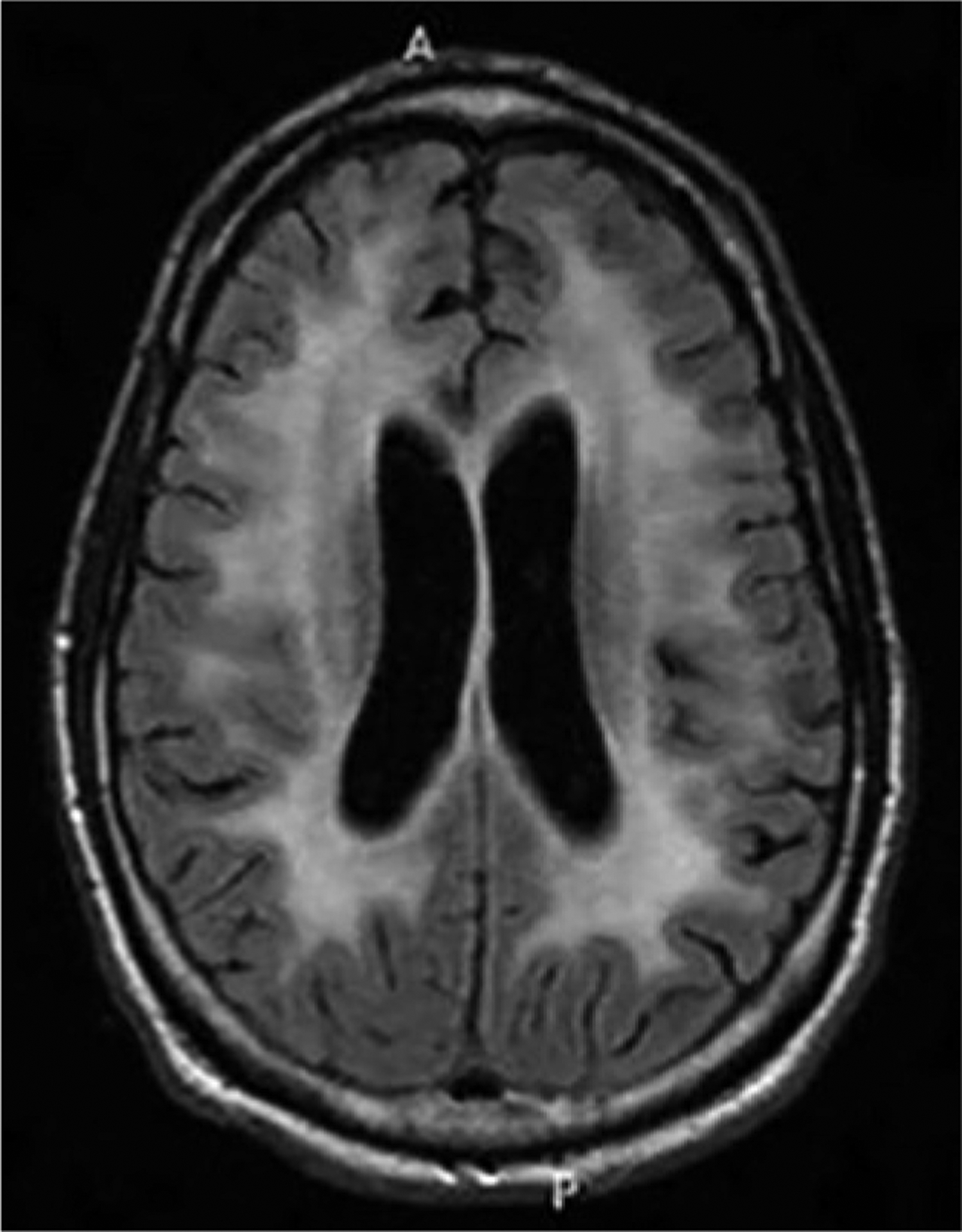
FIGURE 7-2.

Imaging of the patient in CASE 7-1. Axial fluid-attenuated inversion recovery (FLAIR) MRI reveals symmetric confluent subcortical and periventricular white matter hyperintensities without mass effect. Mild cerebral atrophy is also present for the patient’s age.
