
FIGURE 7-4.
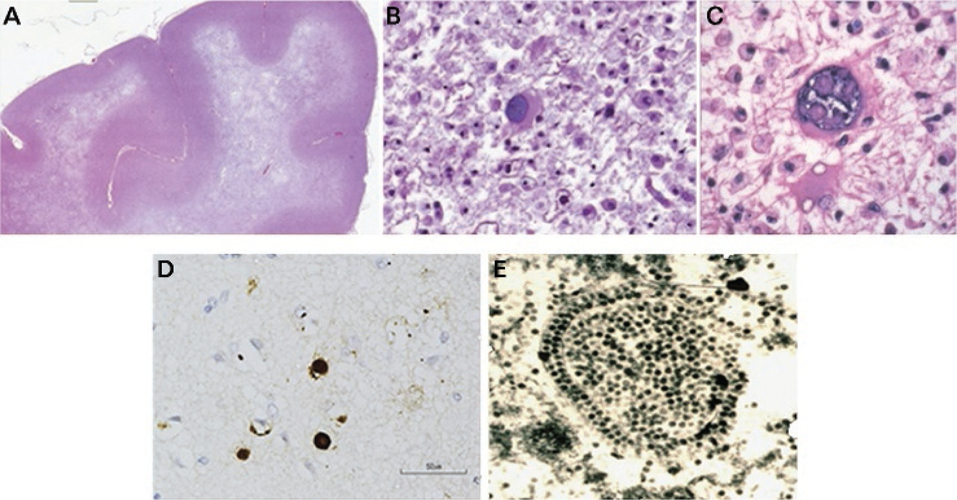
Pathology of progressive multifocal leukoencephalopathy. A, Luxol fast blue staining with hematoxylin counterstain of the frontal lobe in a patient with progressive multifocal leukoencephalopathy (PML) shows extensive multifocal and confluent areas of demyelination. Small islands of demyelination coalesce to produce large confluent areas resulting in a “ground glass” bright appearance on T2-weighted MRI scans. B, Enlarged oligodendrocyte with a large inclusion-bearing nucleus is characteristic of PML. No discrete intranuclear inclusion is seen. C, A large bizarre astrocyte is depicted. D, Immunostaining with polyclonal antibody to JC virus shows dark brown staining of nuclei of several oligodendrocytes. E, Electron micrograph of crystalline array of assembled JC virions in nuclei of infected oligodendrocyte in PML brain lesion. Virions measure 40 nm in diameter.
Reprinted with permission from Berger JR, et al, Neurology.72 © 2013 American Academy of Neurology.