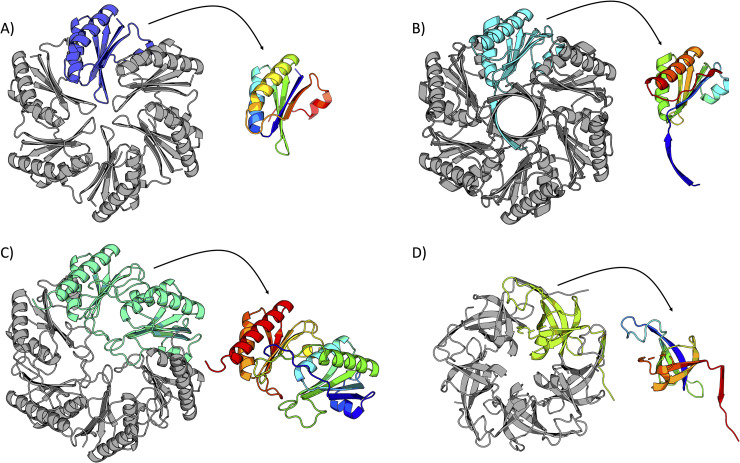
Fig 2. Cartoon representations of four bacterial microcompartment shell proteins.
A single monomer is highlighted and presented in the context of the biological assembly, with a color-ramped (blue = N-terminus; red = C-terminus) version of the monomer adjacent to each structure. (A) A representative hexameric BMC shell protein (BMC-H) (PDB 2EWH) [24]. (B) A representative permuted BMC shell protein (PDB 6XPI) [31]. (C) A representative trimeric BMC shell protein (BMC-T) (PDB 3I82) [28]. (D) A representative BMV shell protein (PDB 4I7A) [15].

