
FIGURE 1:
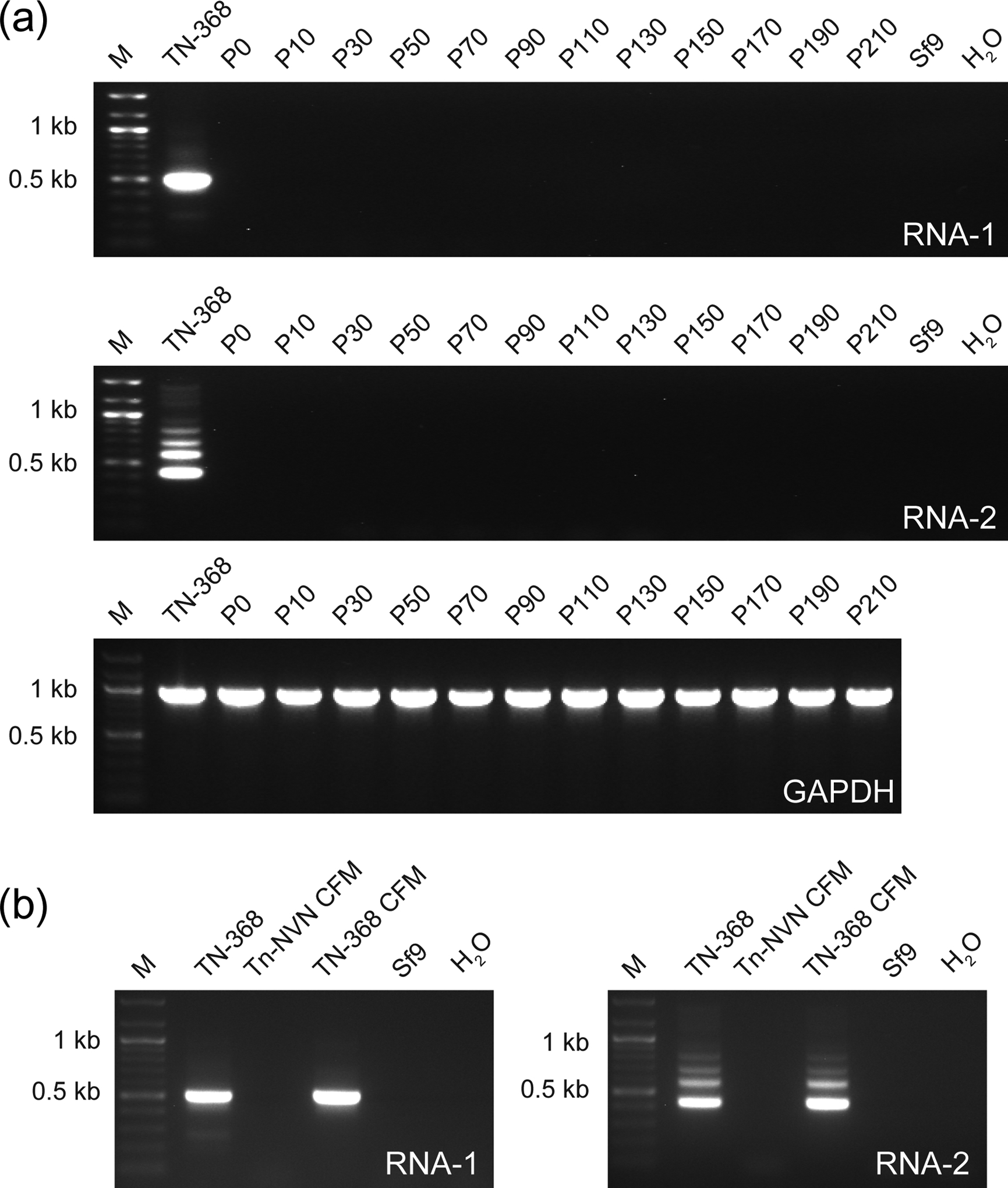
Tn-NVN cells have no detectable TnCLV. (a) A Tn-NVN suspension culture was maintained for 210 serial passages in ESF-921 medium containing no antiviral drugs. Samples were removed periodically and total RNA was isolated from cell pellets and assayed for TnCLV RNA 1, TnCLV RNA 2 by RT-PCR/nested PCR, or Tn GAPDH by RT-PCR. (b) CFM was isolated from a sample taken from the Tn-NVN suspension culture at P55 and ultracentrifuged as described in Materials and methods. Total RNA was then isolated from the resulting high speed pellet and assayed for TnCLV RNA 1 and 2 by RT-PCR/nested PCR. Total RNAs from TN-368 cells and the pellet obtained by ultracentrifuging TN-368 cell-free media were used as positive controls and total RNA from Sf9 cells or PCRs with no RNA template (H2O) were used as negative controls. Each panel in (a) and (b) shows the amplification products obtained using the indicated RNA samples, as determined by agarose gel electrophoresis with ethidium bromide staining. Each lane marked M shows a 100 bp DNA ladder used as a size standard.