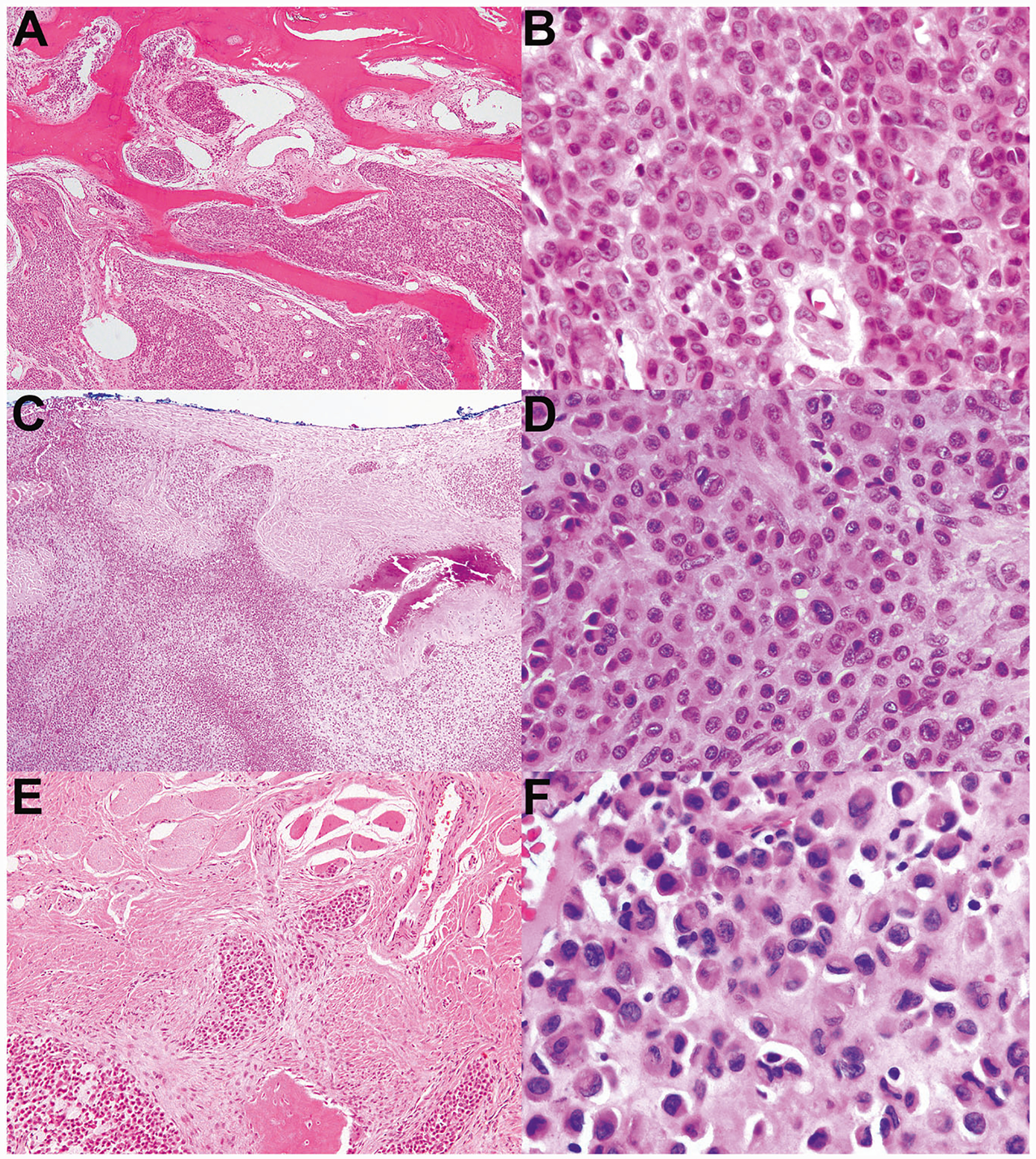
Fig. 4. Malignant chondroblastoma resembling conventional chondroblastoma but with increased atypia and infiltrative borders.

a–f Correspond to cases 2–4, respectively. These three malignant chondroblastomas contained eosinophilic chondroid matrix, numerous admixed osteoclastic giant cells, and neoplastic chondroblasts with eccentric folded nuclei and eosinophilic cytoplasm, all characteristic of conventional chondroblastoma. However, all three exhibited significant cytologic atypia and infiltrative borders, both of which would be unusual for conventional chondroblastoma. a–b This tumor occurred in the talus and had an infiltrative growth pattern. b The degree of cytologic atypia shown here is beyond that of conventional chondroblastoma. c This tumor exhibits a permeative growth pattern; here, it is infiltrating through the cortex of the rib and extending into adjacent soft tissue. d There is significant cytologic atypia, including nuclear pleomorphism and scattered large, hyperchromatic nuclei. e This tumor disrupts the cortex of the rib and extends into adjacent skeletal muscle. f Compared with other malignant chondroblastomas in this series, this tumor had relatively mild nuclear atypia; however, it was found to be markedly infiltrative upon consensus review and was therefore classified as malignant.
