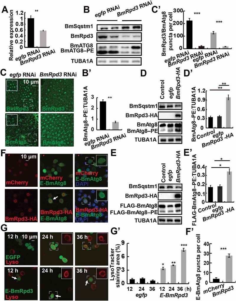
Figure 2.

BmRpd3 is indispensable for autophagy in B. mori. (A-C’) mRNA levels of BmRpd3 (A), protein levels of BmSqstm1, BmAtg8, and BmRpd3 (B), immunofluorescent staining of BmRpd3 and BmAtg8 (C) at 24 h after BmRpd3 RNAi treatment in the fat body. Quantification of BmAtg8–PE in B (B’). Quantification of fluorescent BmRpd3 and BmAtg8 in C (C’). Black columns: BmRpd3, gray columns: BmAtg8–PE, wireframes indicate the magnified fields. (D-D’) Protein levels of BmSqstm1, BmAtg8, and BmRpd3-HA after BmRpd3-HA overexpression for 48 h (D). Quantification of BmAtg8–PE in D (D’), egfp overexpression is used as the control. (E-E’) Protein levels of BmSqstm1, FLAG-BmAtg8, and BmRpd3-HA after co-overexpression of BmRpd3 and FLAG-BmAtg8 for 48 h (E). Quantification of FLAG-BmAtg8–PE in E (E’). (F-F’) Punctation of EGFP-BmAtg8 in BmRpd3 overexpressing cells, mCherry overexpression is used as the control (F). Quantification of BmAtg8 puncta in F (F’). Arrows: the typically treated cells, E-BmAtg8: EGFP-BmAtg8. (G-G’) LysoTracker Red staining after overexpression of egfp-BmRpd3 compared to the egfp-overexpressed control (G). Quantification of LysoTracker Red staining in G (G’), E-BmRpd3: EGFP-BmRpd3, Lyso: LysoTracker Red staining
