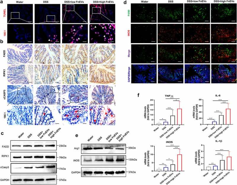
Figure 6.

RIPK1 mediated epithelial cell death drives exacerbated barrier loss in FnEVs-treated colitis mice. (a) Representative images of TUNEL stainings of colon sections on day 3 after colitis induction (red, TUNEL positive; blue, DAPI). Scale bar = 50 um (up) or 20 um (down). (b) Representative images of immunohistochemical stainings of FADD, RIPK1 and cCASP3 in the colon sections on day 3 after colitis induction. Scale bar = 50 um or 20 um (downmost). (c) Immunoblot analysis of protein extracts from colon samples with the indicated antibodies. (d) Double staining of F4/80 (green) and iNOS (red) on day 3 after colitis induction. Nuclear counterstaining is provided with DAPI (blue). Scale bar = 100 um. (e) Immunoblot analysis of protein extracts from colon samples with the indicated antibodies. (f) The relative mRNA level of TNF-α, IL-6, iNOS and IL-1β was detected in colon samples on day 3 after colitis induction. *p < .05, **p < .01, ***p < .001. All data were presented as the means ± SD (n = 6 mice per group)
