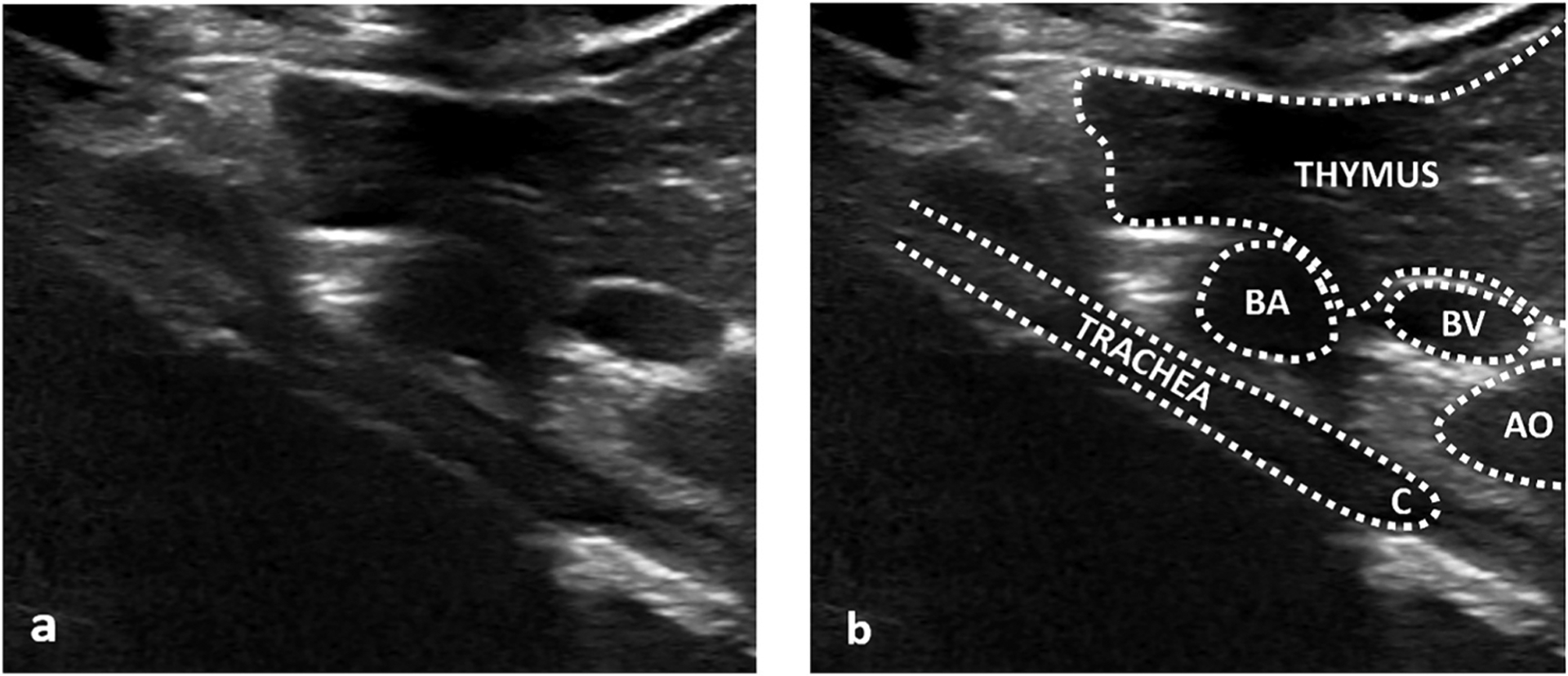
Fig. 1. Tracheal cartilaginous sleeve on ultrasound.

Unlabeled (a) and labeled (b) longitudinal ultrasound images from a right parasternal approach and thymic window showing the abnormal trachea (TRACHEA) as a thick, unsegmented structure in the central mediastinum, ending at the carina (C). Nearby thymus (THYMUS) and vascular structures (BA – brachiocephalic artery, BV – left brachiocephalic vein, AO – aortic arch) are labeled for anatomic orientation. Shadowing deep to the trachea is expected due to the air column. A right parasternal approach is recommended to avoid confusing a gas filled esophagus (not shown) with an unsegmented trachea as their appearance can be similar.
