. 2021 Mar 17;62(4):325–337. doi: 10.3349/ymj.2021.62.4.325

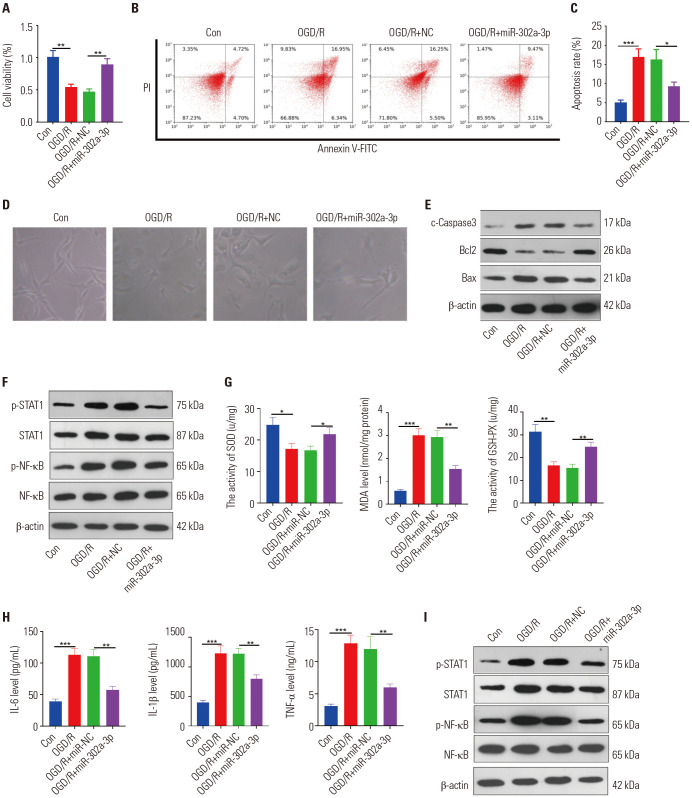
Fig. 4. Overexpressing miR-302a-3p attenuated OGD/R-mediated neuronal damage and microglial activation. The miR-302a-3p mimics were transfected into HT22 neuronal cells and BV2 microglial cells, respectively. (A–C) MTT (A) and flow cytometry (B and C) were performed to determine the proliferation and apoptosis of HT22 cells 24 hours after OGD/R. (D) The image of HT22 cells under different stimulations were observed under a light microscope. (E and F) Western blot was conducted to test the content of Bax, Bcl2, Caspase3, and STAT1/NF-κB proteins in HT22 cells 24 hours after OGD/R. (G) ELISA was conducted to monitor the expressions of SOD, MDA, and GSH-PX in the culture medium of BV2 cells. (H) ELISA was used to detect the levels of IL-6, IL-1β, and TNF-α in the culture medium of BV2 cells. (I) Western blot was performed to examine STAT1 and NF-κB protein content. *p<0.05, **p<0.01, ***p<0.001. n=3. SOD, superoxide dismutase; MDA, malondialdehyde; GSH-PX, glutathione peroxidase; OGD/R, oxygen-glucose deprivation/reperfusion; TNF, tumor necrosis factor; IL, interleukin; ELISA, enzyme-linked immunosorbent assa.

Fig. 4