. 2021 Mar 17;62(4):325–337. doi: 10.3349/ymj.2021.62.4.325

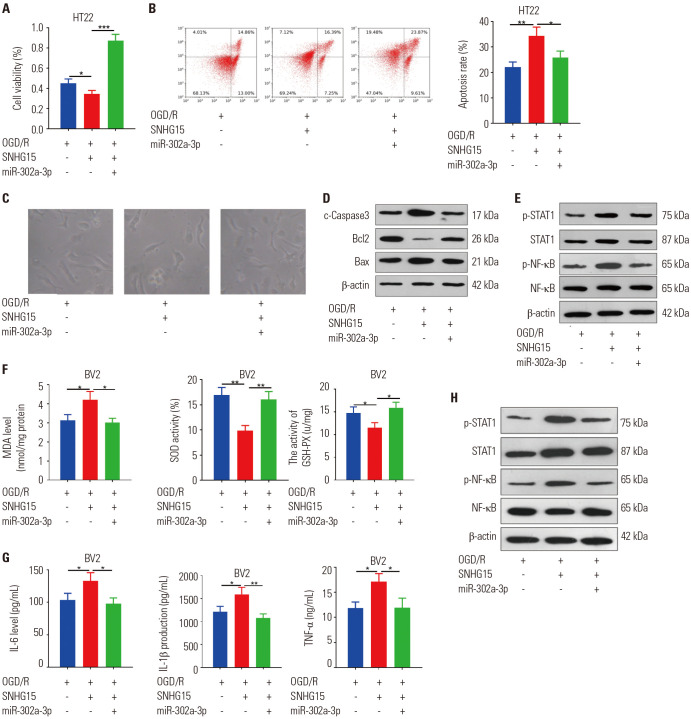
Fig. 6. MiR-302a-3p abated SNHG15-mediated neuronal damage, oxidative stress, and inflammation. The miR-302a-3p mimics were co-transfected with SNHG15 overexpression plasmids into HT22 cells or BV2 cells, which were then treated with OGD/R for 24 hours. (A and B) MTT (A) and flow cytometry (B) were peformed to test HT22 neuronal cell proliferation and apoptosis, respectively. (C) The image of HT22 cells under different stimulations were observed under a light microscope. (D and E) Western blot was employed to determine the content of the apoptosis-related proteins (Bax, Bcl2, and Caspase3) and STAT1/NF-κB pathway in HT22 neuronal cells. (F and G) ELISA was conducted to compare the MDA, SOD, GSH-PX, IL-6, IL-1β, and TNF-α expressions in the culture medium of BV2 cells. (H) Western blot was performed to examine STAT1 and NF-κB protein content. *p<0.05, **p<0.01, ***p<0.001. n=3. OGD/R, oxygen-glucose deprivation/reperfusion; SOD, superoxide dismutase; MDA, malondialdehyde; GSH-PX, glutathione peroxidase; TNF, tumor necrosis factor; IL, interleukin; ELISA, enzyme-linked immunosorbent assay.

Fig. 6