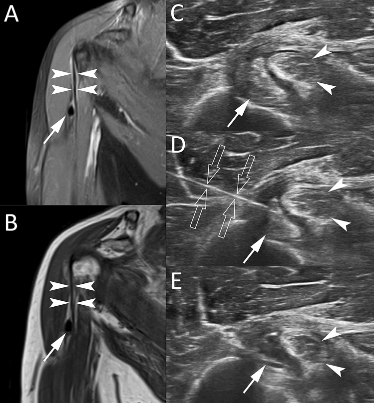
Fig. 10.
Calcific tendinopathy of the long head of the biceps brachii in a 54-year-old male patient. Coronal fat-suppressed proton-density weighted (A) and coronal T1-weighted (B) MR images show a hypointense calcification ovoid in shape (arrows) close to extra-articular portion of the long head of the biceps brachii (headarrows). Short-axis US images (C–E) demonstrate a soft calcification (arrows) within the sheath of the tendon (headarrows). Short-axis US image during the US-PICT (D) shows the needle (void arrows) inside the calcification, which presents anechoic content and a thin calcific wall at the end of the procedure (E)

