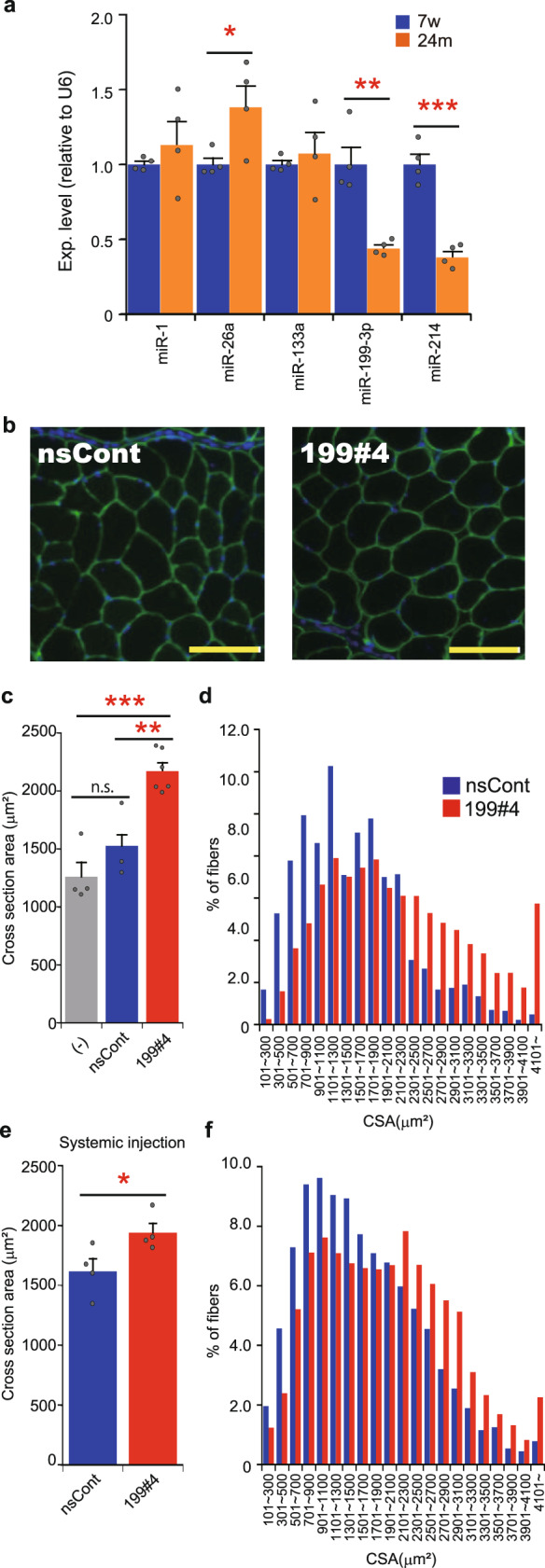
Fig. 5. Effects of miR199#4 on aged mice.

a Expression of miRNAs in young and aged TA muscles. The expression levels of miRNAs (indicated) in the TA muscle of young (7-week-old: 7w) and aged (~24-month-old: 24m) C57BL/6J mice were examined by qRT-PCR and analyzed by the delta–delta Ct method, using the data of U6 snRNA as an internal reference. The data were normalized to the data of young mice as 1. Data are shown as mean ± SEM [n = 4 mice; *p < 0.05, **p < 0.01, ***p < 0.001 by Student’s t test (two-tailed)]. b Photos of the cross-section of myofibers. Aged C57BL/6J mice (~24-month-old) were injected with miR199#4 (199#4) or nsCont to the TA muscle, and 2 days later the TA muscles were examined as in Fig. 4b. Scale bars indicate 100 µm. c Averaged cross-sectional area of myofibers. The cross-sectional areas (CSAs) of myofibers in the TA muscle were measured and analyzed as in Fig. 4d. The CSAs of myofibers in untreated aged mice (−) were also examined. The number of mice examined was as follows: six miR199#4-dosed mice, three nsCont-dosed mice, and four untreated mice. Data are shown as mean ± SEM [**p < 0.01, ***p < 0.001 by ANOVA (Tukey’s post hoc test)]. d Histogram of the CSAs of myofibers. Histogram of the CSAs of myofibers was analyzed and displayed as in Fig. 4e. e, f Analyses of the CSAs of myofibers in aged mice intravenously injected with miR199#4. Aged C57BL/6J mice (~24-month-old) were intravenously injected with miR199#4 or nsCont from the tail vein. One week later, the CSAs of myofibers in the TA muscle were examined as in Fig. 4b; and averaged CSA (e) and histogram of the CSAs (f) were analyzed as in Fig. 4d, e, respectively. The data of averaged CSAs (e) are shown as mean ± SEM [n = 4 treated mice; *p < 0.05 by Student’s t test (two-tailed)].
