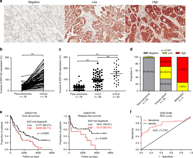
Fig. 1. Expression of EEF1A2 and clinical significance in lung adenocarcinoma (LUAD).
EEF1A2 expression was detected by immunohistochemistry (IHC) of paraffin-embedded tissues of LUAD patients. a Representative images were shown for negative, low or high expression of EEF1A2 staining. b Quantitative analysis of EEF1A2 expression in 78 paired LUAD tissues (T) and adjacent tissues (N). c EEF1A2 expression was positively related to the clinical pathologic stage (Para-carcinoma, n = 78; I, I–II, II n = 58; II–III, III n = 20). d The distribution of EEF1A2 expression levels in different tissues. (para-carcinoma n = 78, primary lesion n = 78 and metastasis n = 4). e Kaplan–Meier curve analysis of overall survival (OS) and relapse-free survival (RFS) of LUAD patients according to different EEF1A2 expression levels. f Receiver operating characteristic (ROC) curves of EEF1A2 for the diagnosis of LUAD. (Healthy, n = 65; LUAD patients, n = 45). (AUC = 0.7761, 95% CI: 0.6778 to 0.8743) *p < 0.05, **p < 0.01, and ***p < 0.001, NS no statistical significance.

