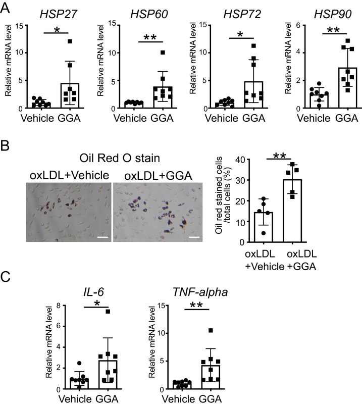
Figure 5.
Effect of the induction of heat shock proteins (HSPs) on peritoneal macrophages. (A) HSP27, 60, 72, and 90 mRNA expression levels in ApoE−/− peritoneal macrophages after geranylgeranylacetone (GGA) treatment. Peritoneal macrophages were isolated from 8-week-old ApoE−/− mice. After the cells were treated with vehicle or GGA (10 μM) for 24 h, total RNA was extracted. (B) Representative micrographs of Oil Red O staining of peritoneal macrophages (left). Scale bar = 100 µm. Vehicle or GGA (10 μM) was added to the cells, and oxidized low-density lipoproteins (oxLDL) (100 μg/mL) were administered 24 h later. One day later, the cells were fixed and stained. Bar graph quantifying Oil Red O positive cells in peritoneal macrophages (right). (C) Interleukin 6 (IL-6) and tumor necrosis factor-alpha (TNF-alpha) mRNA expression levels in ApoE−/− peritoneal macrophages after GGA treatment. Peritoneal macrophages were isolated from 8-week-old ApoE−/− mice. After the cells were treated with vehicle or GGA (10 μM) for 24 h, total RNA was extracted. Each bar represents the mean ± standard deviation (SD). Significant differences are marked on the chart (*p < 0.05; **p < 0.01). n = 5–8 per group.

