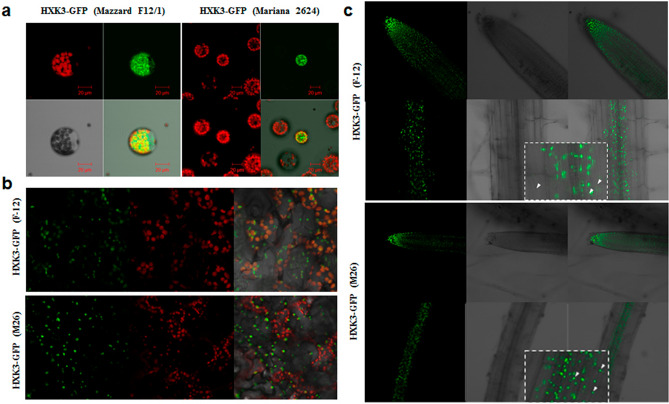
Figure 3.
Subcellular localization of Prunus HXK3-GFP proteins. (a) Transient expression in Arabidopsis thaliana protoplasts. (b) Stable expression in Arabidopsis leaves. (c) Stable expression in Arabidopsis roots. Cells rich in chloroplast as epidermal pavement and mesophyll cells were detected by chlorophyll fluorescence (red) and HXK3-GFP was detected by green fluorescence. After transformation, the samples were incubated for 24 h at 22 °C (Arabidopsis protoplasts). The samples were visualized by confocal microscope. The experiments were done in triplicate, resulting with the same fluorescence patterns.

