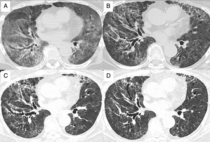
Figure 1.

Computed tomography images of the chest (A) when requiring mechanical ventilation; (B) two weeks after extubation, requiring high‐flow nasal cannula oxygen therapy and nasogastric tube feeding; (C) when initiating nintedanib therapy; and (D) two months after the implementation of nintedanib treatment.
