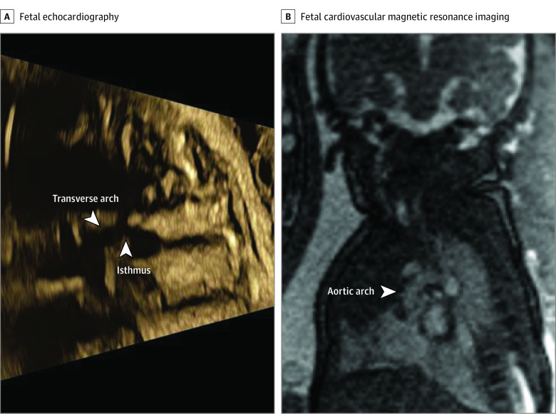
Figure 2. Evaluation of Aortic Arch Anatomy in a Fetus With Suspected Aortic Arch Hypoplasia and/or Coarctation.
Fetal echocardiography (A) showed suspected aortic arch hypoplasia and/or coarctation. The arrows indicate a narrow transverse aortic arch and aortic isthmus. However, fetal cardiovascular magnetic resonance imaging showed a normal-sized aortic arch with no signs of coarctation (B). The CMR findings were confirmed by postnatal echocardiography (not shown). Contrast and brightness in the CMR image have been optimized for printing (case 8; eTable in the Supplement).

