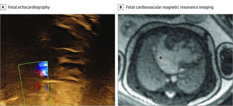
Figure 4. Evaluation of Atrial Restriction in a Fetus With Hypoplastic Left Heart Syndrome.
Fetal echocardiography could not visualize the atrial cavity or Doppler flow across the atrial septum due to poor acoustic windows (A). Fetal cardiovascular magnetic resonance showed a large interatrial communication, indicated by the asterisk, and no nutmeg pattern (B). Therefore, the risk of restrictive atrial septum was considered low (albeit a membrane could not be ruled out), and the fetus was planned for a vaginal delivery without cardiac catheterization laboratory on standby. Contrast and brightness in the CMR image have been optimized for printing (case 5) (eTable in the Supplement).

