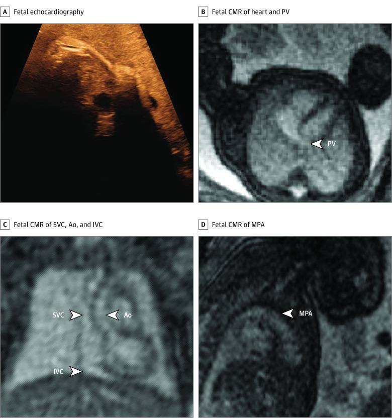
Figure 5. Basic Assessment in a Fetus With Risk Factors for Cardiac Malformation and a Very Poor Acoustic Window.
The fetal heart and vessels were not visible at all during fetal echocardiography, due to mother having obesity (A). Fetal cardiovascular magnetic resonance imaging (MRI) showed normal-sized ventricles with normal systolic function and at least 1 normal pulmonary vein (PV) without obvious direct or indirect signs of aberrant pulmonary veins (B), superior venae cavae (SVC) and inferior venae cavae (IVC) and normal ascending aorta (Ao) (C), and normal main pulmonary artery (MPA) (D). This information ruled out major congenital heart disease, and delivery was planned at the hospital closest to the patient’s home. A nonurgent postnatal echocardiography examination was performed before discharge, which showed a normal cardiovascular anatomy. Contrast and brightness in the CMR images have been optimized for printing (case 30) (eTable in the Supplement).

