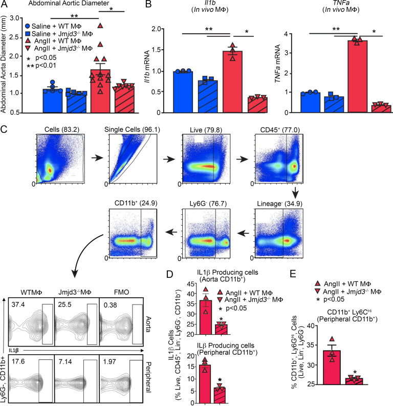
Figure 6.
AAA formation is inhibited in macrophage-specific JMJD3-deficient mice. (A) Maximal abdominal aortic diameter was determined by ultrasound in Jmjd3−/−MΦ or WTMΦ mice infused with either saline or AngII (n = 5 in saline-infused cohorts and 7–10 in AngII-infused cohort). *, P < 0.05; **, P < 0.001, ANOVA with Newman–Keuls multiple comparison test. (B) Quantitative PCR analysis of Il1b and Tnfa from macrophages (CD11b+[CD3−CD19−Nk1.1−Ly6G−]; n = 3 mice/group run in triplicate). *, P < 0.05; **, P < 0.01, ANOVA test with Tukey's multiple comparison test. (C) Gating strategy to select single, live, lineage− [CD3, CD19, NK1.1, Ter-119]−, Ly6G−, CD11b+ by flow cytometry at day 28 peripheral blood monocytes and aortic tissue macrophages. (D) Percentage of CD11b+ cells from monocytes and aortic tissue macrophages immunostained positive for IL-1β in Jmjd3−/−MΦ or WTMΦ mice (n = 3/group run in triplicate). Tissues of two mice per mouse were pooled for a single biological replicate. *, P < 0.05 by Welch’s t test. (E) Percentage of CD11b+Ly6CHi cells in Jmjd3−/−MΦ or WTMΦ mice (n = 3 mice/group). *, P < 0.05 by Welch’s t test. Data are presented as the mean ± SEM. MΦ, macrophage.

