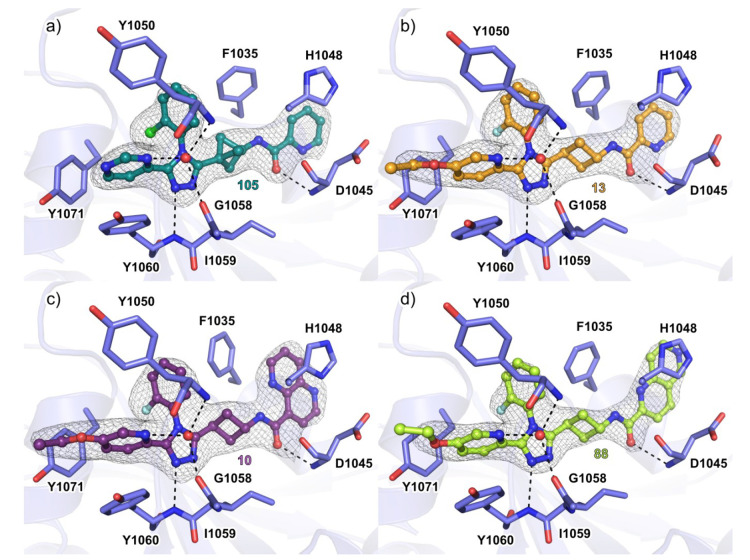
Figure 3.
Cocrystal structures of TNKS2 with inhibitors. (a) Binding mode of 105 with TNKS2 catalytic domain (PDB code 6TKN). (b) Binding mode of 13 with TNKS2 catalytic domain (PDB code 6TG4). (c) Binding mode of 10 with TNKS2 catalytic domain (PDB code 6TKM). (d) Binding mode of 88 with TNKS2 catalytic domain (PDB code 6TKS). The dashed lines in black represent hydrogen bonds, and the red spheres represent water molecules. The σA weighted 2Fo – Fc electron density maps around the ligands are contoured at (1.4–1.7)σ. Structures were solved with molecular replacement using the structure of TNKS2 (PDB code 5NOB) as a starting model.

