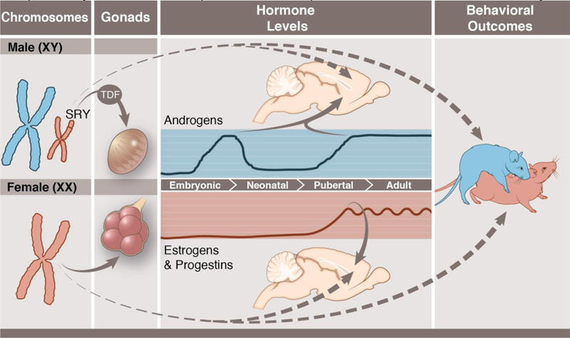
Figure 1: Sexual differentiation of the brain and behavior.

Male typical (top; blue) and female typical (bottom; pink) sexual differentiation. Sex chromosome complement (XX in females; XY in males) leads to sex determination. The Y-chromosome gene, SRY, codes for the protein testicular determining factor (TDF), which programs the bi-potential gonads to differentiate into testes (left). In rodents, the testes begin secreting androgens during the late embryonic period, and these androgens lead to sexual differentiation of the brain and other tissues (middle). In the rodent brain, androgens are converted into estrogens, and estrogens masculinize the brain. In genetic females, the gonads differentiate into ovaries, and the ovaries are largely quiescent in the perinatal period (middle), thus brain feminization occurs largely in the absence of active hormonal signals. At puberty, both male and female gonads produce sex-typical hormone secretions, namely high androgen levels and high but pulsatile estrogen and progestin levels in females. Both early life (organizational) and post-pubertal (activational) hormones contribute to sex differences in behavior, most notably male mounting behavior (blue rat) and female lordosis behavior (pink rat) (right). Sex chromosomes can also have independent effects on brain organization as well as adult sex differences in behaviors (dashed lines). Anthony S. Baker, CMI, Reproduced with the permission of The Ohio State University.
