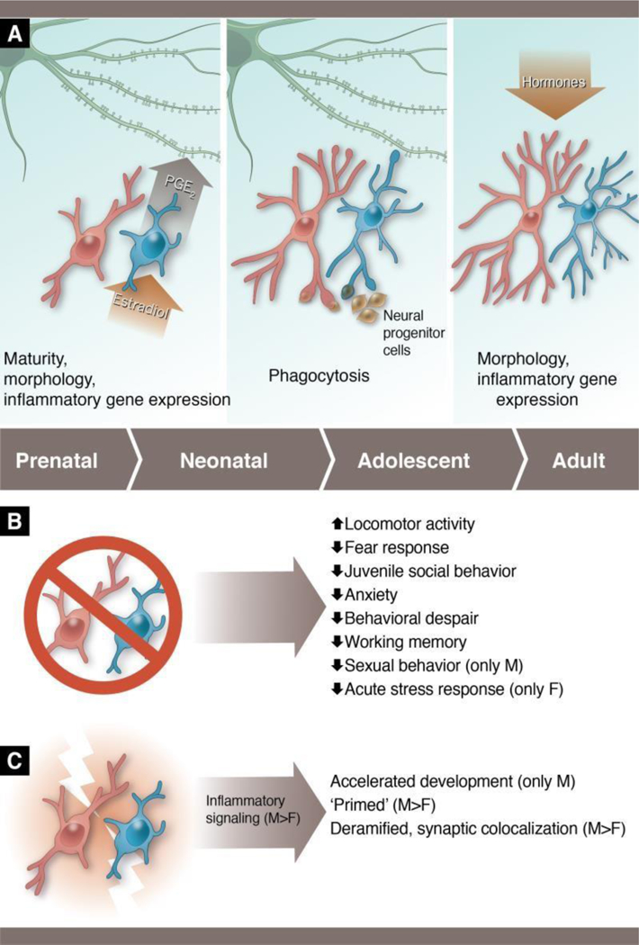
Figure 2: Sex differences in microglia across different developmental windows and conditions.

(A) During the perinatal period (left panel), males have more microglia than females, and the microglia are in a more activated and immature form. Microglia in the male preoptic area respond to the masculinizing hormone, estradiol, by releasing the inflammatory lipid, prostaglandin E2 (PGE2), and PGE2 is responsible for programming male-typical dendritic spine genesis/maintenance that is necessary to display adult copulatory behavior. During the perinatal-juvenile period (middle panel), female microglia appear to show a higher level of phagocytic activity in the hippocampus than male microglia. Female microglia target neural progenitor cells at a higher rate than males, and higher phagocytosis in females is also correlated with reduced dendritic spines in the hippocampus at this time, though they have not been causally linked to date. In adulthood (right), female microglia are hormone responsive and show hyper-ramification relative to males. (B) Studies in which microglia have been depleted early in life demonstrate lifelong behavioral changes in both males and females, with sex-specific effects being seen in some outcomes. (C) Studies in which microglia are stimulated early in life show that male microglia are more responsive than female microglia. Male microglia depicted in blue and female microglia in pink throughout. Figure based on data from Bollinger et al., 2016; 2017; Hanamsagar et al., 2017; Hui et al., 2018; Lenz et al., 2013; Makinson et al., 2017; Nelson & Lenz, 2016; Nelson et al., 2017; Schwarz et al., 2011; Van Ryzin et al., 2016, Weinhard et al., 2018b). Anthony S. Baker, CMI, Reproduced with the permission of The Ohio State University.
