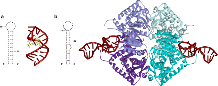
Fig. 8.
Aptamer structures and their interactions with the targets. a The 2D structure of RNA aptamer recognizing an aminoglycoside antibiotic, neomycin B, and the 3D structure of the aptamer (red) bound to the target (yellow) (PDB accession number: 1NEM). The stem of the RNA aptamer forms a pocket with the three consecutive mismatches and parts of the loop interacting with the target. b The 2D model of 2008s ssDNA aptamer and 3D representation of two aptamers (red) bound to tetramer complex of Plasmodium falciparum lactate dehydrogenase (light and dark lilac and light and dark cyan) (PDB accession number: 3ZH2). The 2008s aptamer is able to recognize two distinct sites on the dehydrogenase molecule via interaction with different nucleotides. The 2D aptamer structures were generated using mfold web server and the 3D structures bound to the specific target were modified in Discovery Studio Visualizer

