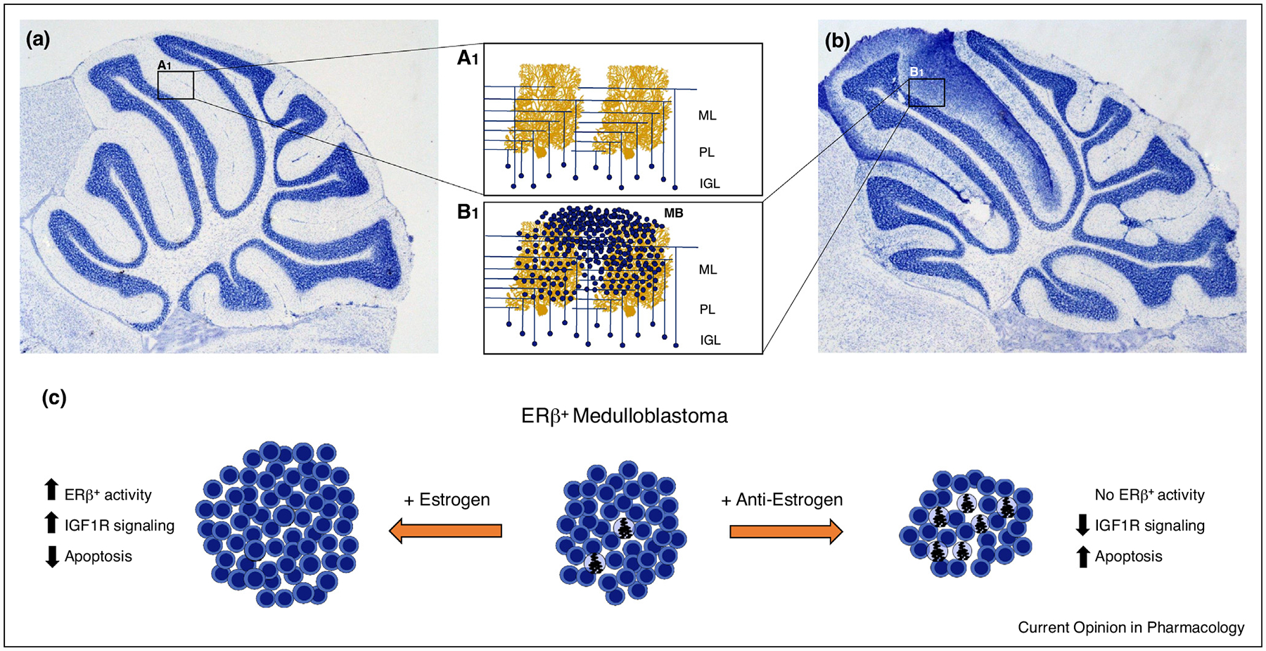
Figure 2.

ERβ activation drives progression of medulloblastoma through cytoprotective and anti-apoptotic mechanisms. Granule cells in the IGL of the healthy mouse cerebellum (a) only weakly express ERβ, leading to a highly structured network of mossy fiber and parallel fiber synapses on Purkinje cell dendrites [A1]. Dysregulation of granule cell mitogenesis results in cerebellar overgrowth, excessive granule cell numbers in the IGL and tumor formation associated with the ML (b). In MB cells (c), ERβ is highly expressed and estrogen drives tumor formation and progression through cytoprotective mechanisms that decrease apoptosis. Addition of anti-estrogens (+ anti-estrogen) block the protective ERβ actions increases apoptosis resulting in decreased tumor size. Abbreviations: ML, molecular layer; PL, Purkinje cell layer; IGL, internal granular layer. Purkinje cell adapted from Cell Image Library CCDB_3687.
