Figure 3.

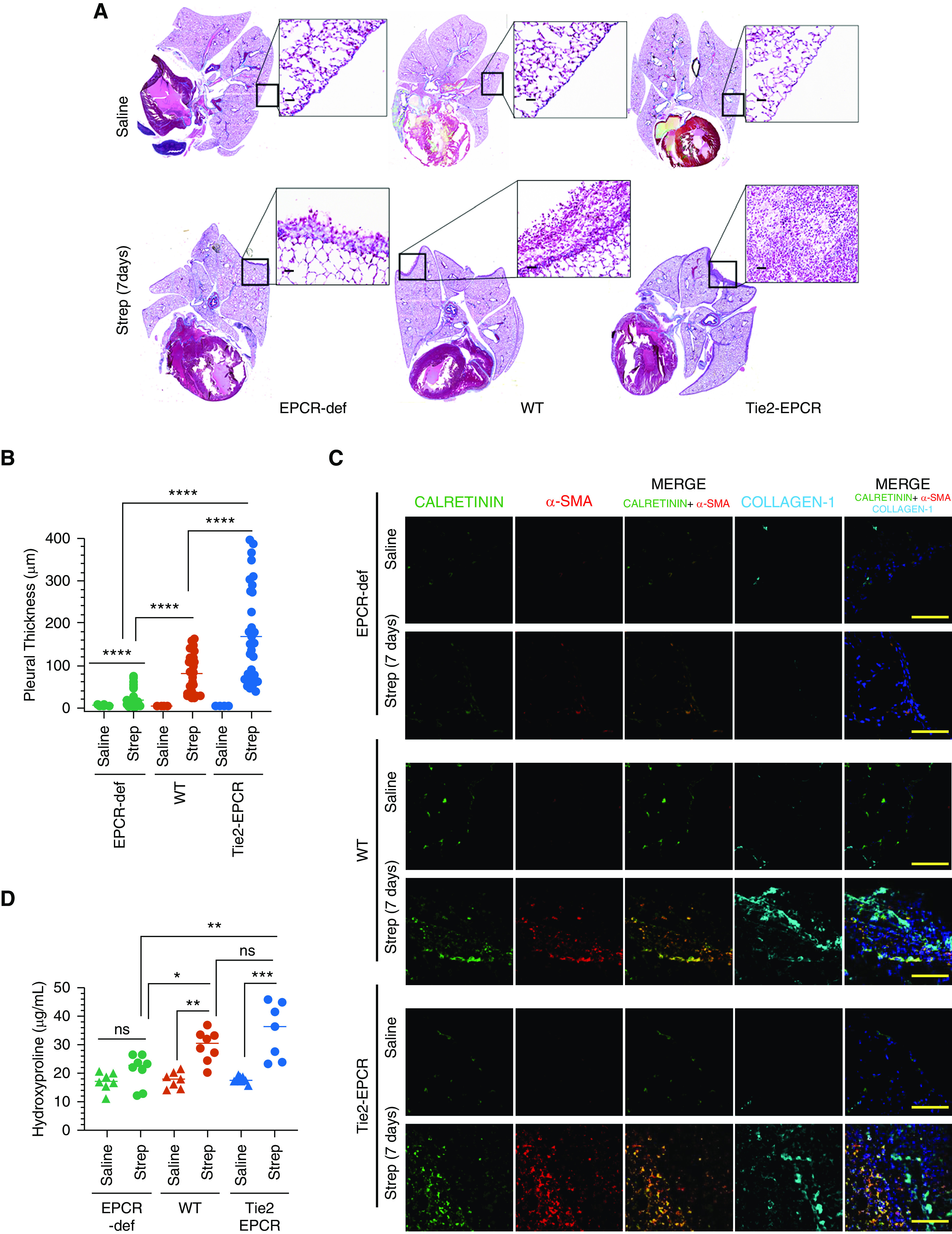
EPCR promotes pleural thickening induced by S. pneumoniae infection. WT, EPCR-overexpressing Tie2-EPCR, or EPCR-def mice were administered with saline or S. pneumoniae intrapleural. The lungs were harvested 7 days after infection. (A) Masons-Trichrome staining of lung tissue sections to assess collagen deposition. (B) Pleural thickening, as measured microscopically at random places covering the entire lungs using Nikon digital sight DS-Fi1 camera (Nikon Instruments) and NIS elements BR 3.2 (Nikon Instruments) software (number of fields examined per group, 34–44). (C) Immunofluorescence staining of lung tissue sections from saline-administered or S. pneumoniae–infected mice for calretinin (as a mesothelial marker), α-smooth muscle actin (α-SMA), and collagen-1. The images of Calretinin were merged with the images of α-SMA (middle column) or α-SMA+collagen (last column on the right). Scale bars, 100 µm. (D) Hydroxyproline content in the lung tissues of saline and S. pneumoniae–infected mice (7 d). Scale bars, 20 µm. *P < 0.05, **P < 0.01, ***P < 0.001, and ****P < 0.0001.
