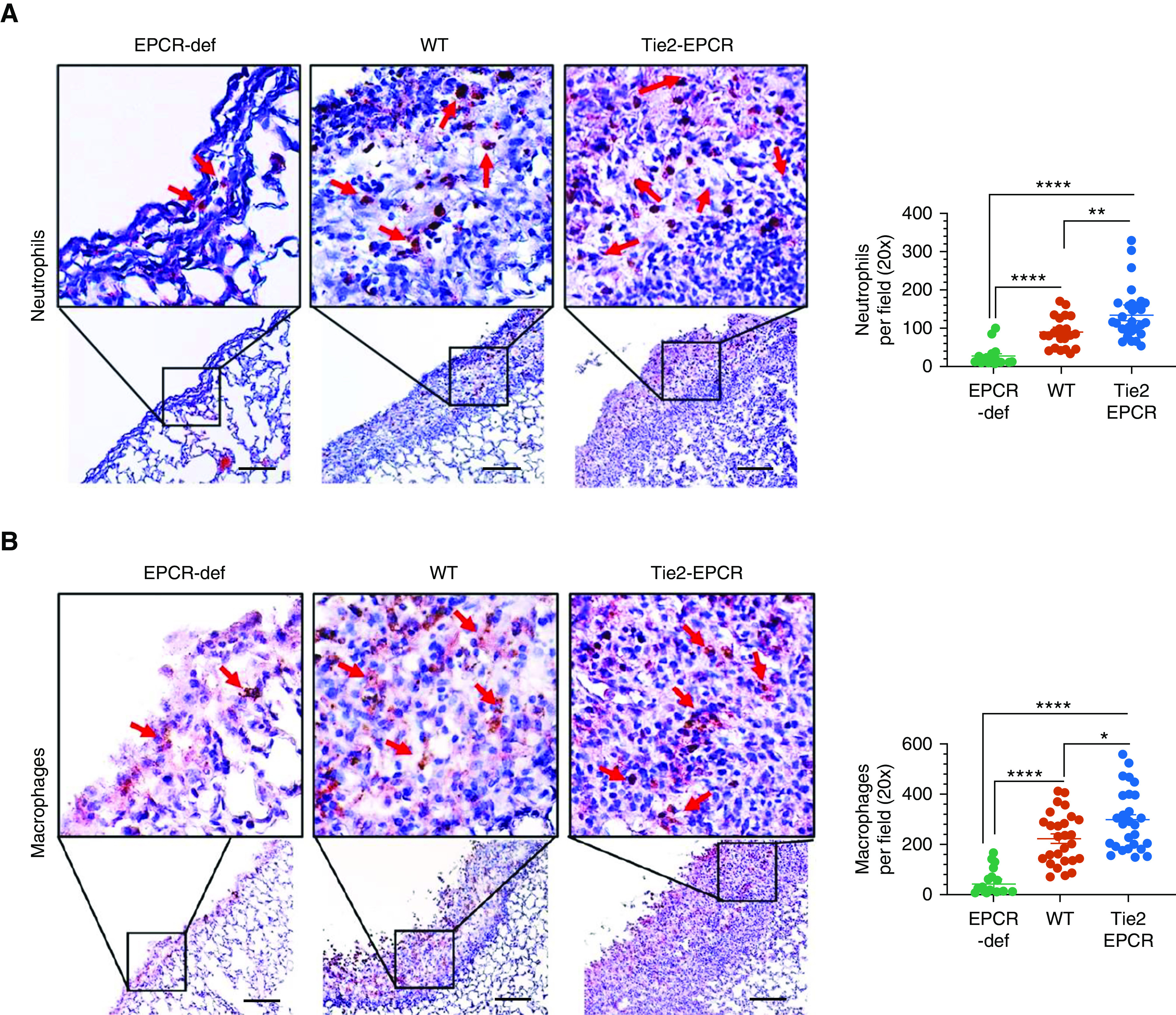
Figure 4.

Increased infiltration of neutrophils and macrophages into the fibrotic pleura in EPCR-expressing mice after S. pneumoniae pleural infection. WT, Tie2-EPCR, or EPCR-def mice were administered with saline or S. pneumoniae intrapleural. After 7 days, lungs and pleural lavages were harvested from these mice. The lung tissue sections were stained with antimouse Ly6G to identify neutrophils (n = 15–28 fields per group) (A) or with anti-F4/80 to mark macrophages (n = 19–25 fields per group) (B). The number of neutrophils and macrophages in multiple fields was counted and averaged. These data were shown in the scatter dot plot graph, the right side of the images. Red arrows point to neutrophils or macrophages. Scale bars, 100 µm. *P < 0.05, **P < 0.01, and ****P < 0.0001.
