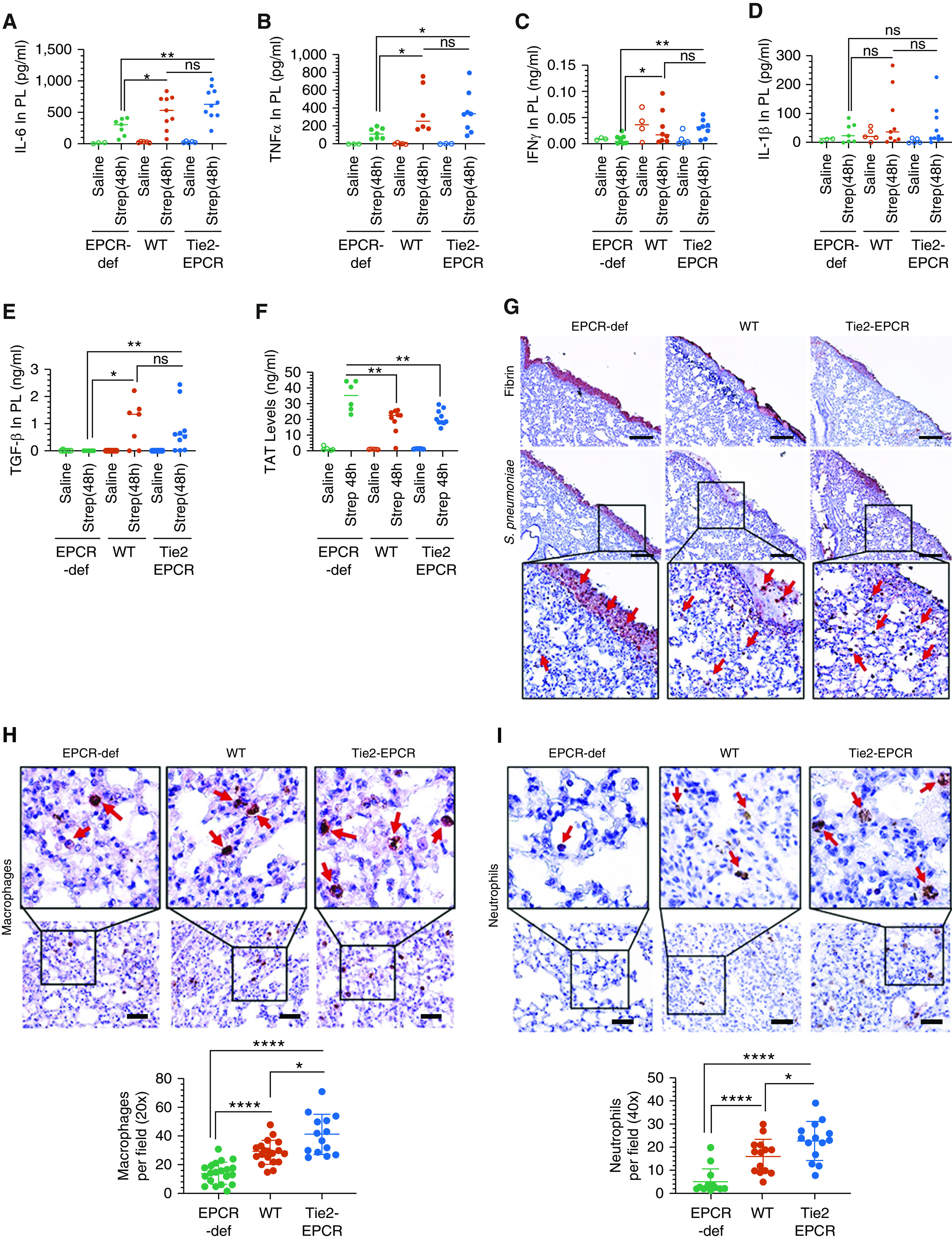
Figure 5.

EPCR-def mice exhibit reduced inflammation but enhanced coagulation and pleural fibrin deposition. Saline or S. pneumoniae was administered into the pleural cavity of WT, Tie2-EPCR, or EPCR-def mice. After 48 hours, mice were killed, and pleural lavages and tissues were collected. (A–E) Pleural lavages (PL) were analyzed for the presence of proinflammatory cytokines IL-6 (A), TNF-α (B), IFN-γ (C), IL-1β (D), and TGF-β (E). (F) Thrombin-antithrombin complex levels were also estimated in the pleural lavages (saline, n = 3–5 mice per group; S. pneumoniae infection, n = 7–10 mice per group). (G–I) Lung tissue sections were stained by immunohistochemistry for fibrin (G, top panel) and S. pneumoniae (type 2 serotype) (G, bottom panel, insets were enlarged digitally), macrophages (F4/80 antigen) (H), and neutrophils (Ly6G antigen) (I). Macrophages and neutrophils number was counted from 14 to 20 fields from tissue sections originated from three to five mice/group. Red arrows point to (G) S. pneumoniae, (H) macrophages, or (I) neutrophils. Scale bars, 100 µm. *P < 0.05, **P < 0.01, and ****P < 0.0001.
