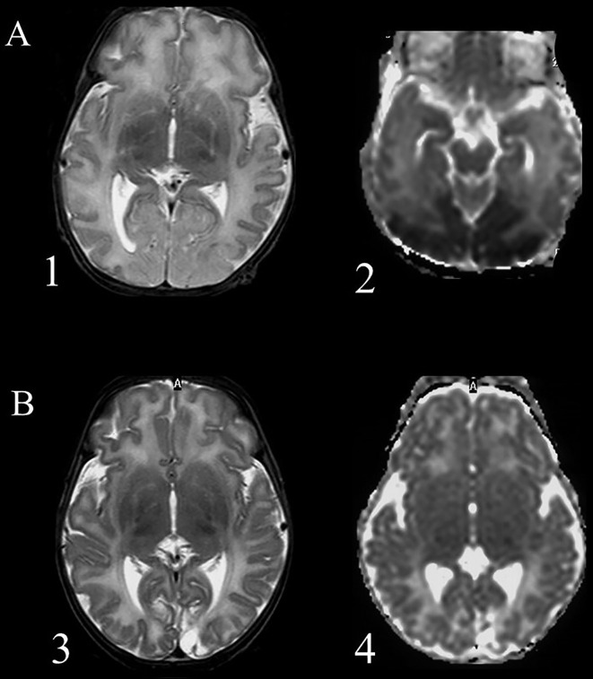
Figure 1.
Magnetic resonance imaging of a term newborn with apnea and seizures associated with severe hypoglycemia. (A) scans performed after 5 days from the event (acute phase), 1 = axial T2 weighted imaging showing an extensive cortical and subcortical damage, manly in the occipital lobes, with loss of cortical differentiation; 2 = Diffusion weighted imaging shows a large posterior subcortical area of reduction of the apparent diffusion coefficient. (B) scans performed after 5 weeks (chronic phase): 3 = Presence of an occipital atrophic area, more extended within the left occipital lobe; 4 = Diffusion weighted imaging shows an increased water diffusion in the affected areas and a smaller cavitation.

