
Figure 5.
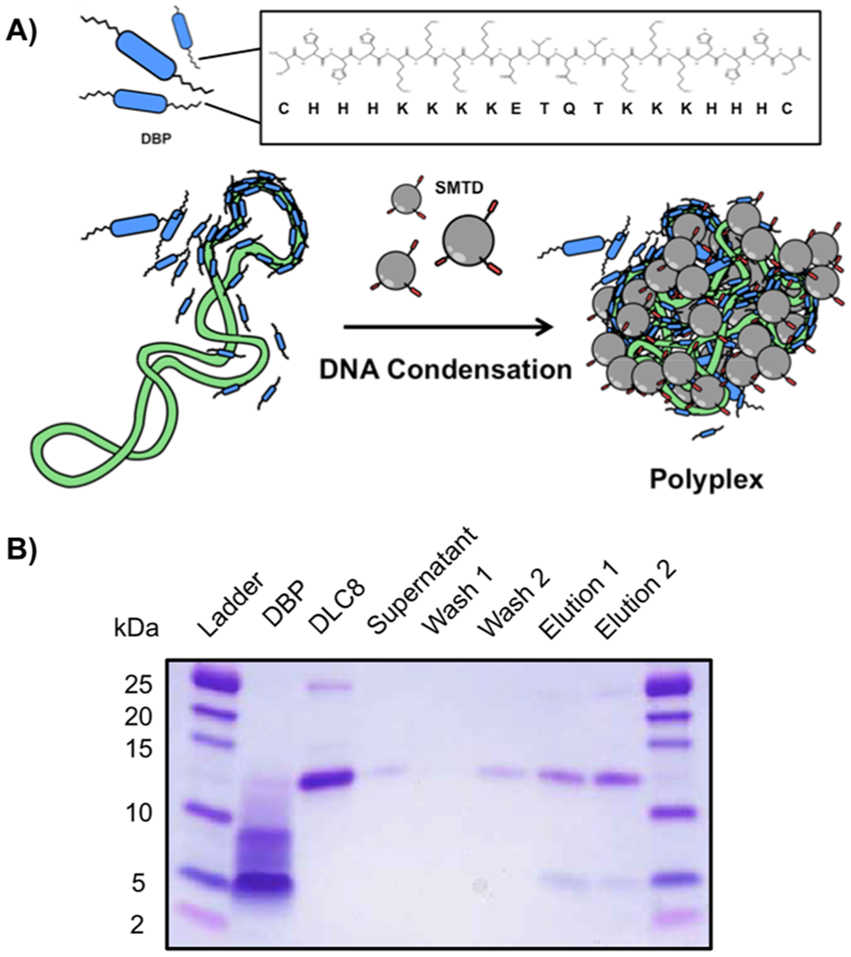
Association of DBP with DLC8. (A) Schematic illustrating the formation of the polyplexes with DBP, G5-SMTP, and plasmid DNA. DBP is in blue, the plasmid DNA is in green and the SMTP is in gray. The primary sequence of DBP is also shown in the inset. (B) SDS-PAGE analysis of the in vitro binding pull-down assay between DBP and DLC-8.