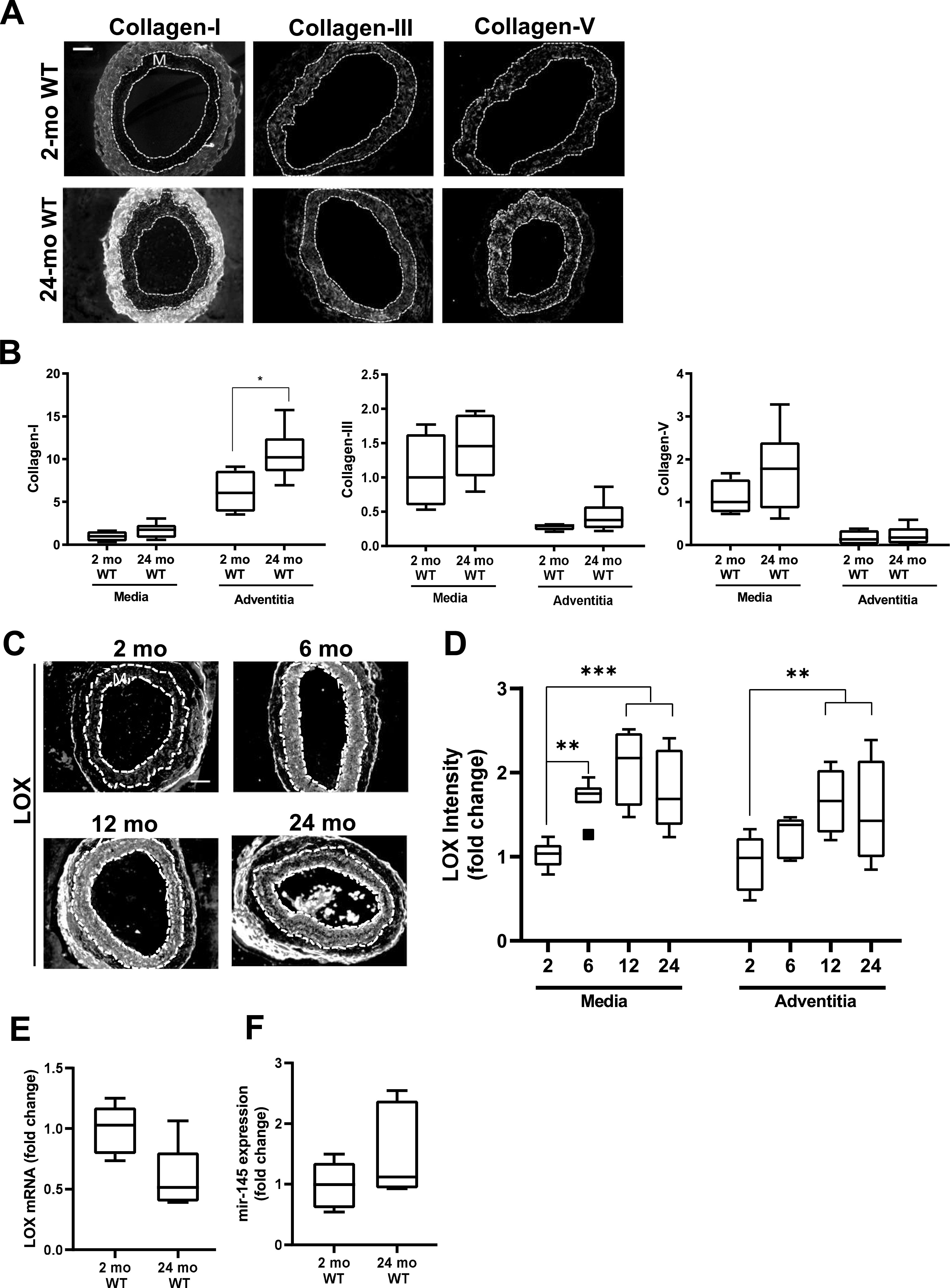
Figure 8. Differential mechanisms drive overexpression of lysyl oxidase (LOX) in normal aging.

C57BL/6 (WT) mice were aged from 2 to 24 mo. (A) Representative images of collagen-I, collagen-III, collagen-V immunostaining in carotid artery sections of 2- and 24-mo WT mice (n = 4–7 mice per age-group). (B) Collagen signal intensities in the medial and adventitial layers of the immunostained carotid cross sections were quantified, and results were normalized to the mean of the 2-mo WT medial layer for each collagen. Statistical significance was determined by Mann–Whitney tests between ages. (C) Representative images of LOX immunostaining of carotid artery cross sections from 2-mo (n = 8), 6-mo (n = 7), 12-mo (n = 4), and 24-mo (n = 9) WT mice. (D) LOX signal intensities were quantified from the immunostained sections, and results were normalized to the mean signal intensity of the 2-mo WT medial layer. Statistical significance was determined by one-way ANOVA relative to the 2-mo mice followed by Holm–Sidak post-tests. (E, F) LOX mRNA (n = 5 per age) and (F) miR-145 expression levels (n = 6 per age) in 2- and 24-mo WT aorta determined by RT-qPCR and normalized to 2-mo WT. Statistical significance was determined using Mann–Whitney tests. In carotid cross section images, the arterial media (M) is outlined with dashed lines; scale bar = 50 μm.
